

Fred Ruhe
Well-known member

Anusuya Chinsamy, Delphine Angst, Aurore Canoville, & Ursula B. Göhlich, in press.
Bone histology yields insights into the biology of the extinct elephant birds (Aepyornithidae) from Madagascar
Biological Journal of the Linnean Society. in press. doi:10.1093/biolinnean/blaa013
Abstract: https://academic.oup.com/biolinnean...nnean/blaa013/5815707?redirectedFrom=fulltext
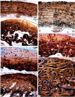
Given that the biology of the recently extinct aepyornithids is poorly understood, we undertook a histological study of 29 skeletal elements of adult and juvenile specimens of Aepyornithidae, i.e. Aepyornis maximus, Aepyornis hildebrandti and Vorombe titan, in addition to a group of taxonomically unidentifiable juvenile Aepyornithiformes. Comparative analysis of the histology of the different skeletal elements showed that although the femur retained a good record of growth during early ontogeny, the tibiotarsus provided the best record of growth. Our data showed that, like other insular birds and their extant relative, the kiwi, Aepyornithidae experienced protracted growth. We also found that intracortical remodelling began early in ontogeny and continued throughout their lives, becoming more extensive throughout the compacta with age, albeit more restricted to the perimedullary region in the femora. We also deduced that the different skeletal elements experienced variable amounts of intracortical remodelling, which was most likely to be related to biomechanical constraints, size of the element and ontogenetic age. Additionally, we documented the occurrence of an unusual endosteal tissue within the large perimedullary erosional spaces of a femur of A. maximus. Overall, our study provided a lot of new information about the life history of these giant, recently extinct ratites.
Enjoy,
Fred
Bone histology yields insights into the biology of the extinct elephant birds (Aepyornithidae) from Madagascar
Biological Journal of the Linnean Society. in press. doi:10.1093/biolinnean/blaa013
Abstract: https://academic.oup.com/biolinnean...nnean/blaa013/5815707?redirectedFrom=fulltext
Given that the biology of the recently extinct aepyornithids is poorly understood, we undertook a histological study of 29 skeletal elements of adult and juvenile specimens of Aepyornithidae, i.e. Aepyornis maximus, Aepyornis hildebrandti and Vorombe titan, in addition to a group of taxonomically unidentifiable juvenile Aepyornithiformes. Comparative analysis of the histology of the different skeletal elements showed that although the femur retained a good record of growth during early ontogeny, the tibiotarsus provided the best record of growth. Our data showed that, like other insular birds and their extant relative, the kiwi, Aepyornithidae experienced protracted growth. We also found that intracortical remodelling began early in ontogeny and continued throughout their lives, becoming more extensive throughout the compacta with age, albeit more restricted to the perimedullary region in the femora. We also deduced that the different skeletal elements experienced variable amounts of intracortical remodelling, which was most likely to be related to biomechanical constraints, size of the element and ontogenetic age. Additionally, we documented the occurrence of an unusual endosteal tissue within the large perimedullary erosional spaces of a femur of A. maximus. Overall, our study provided a lot of new information about the life history of these giant, recently extinct ratites.
Enjoy,
Fred
Last edited: