

Fred Ruhe
Well-known member

Anusuya Chinsamy and Trevor H. Worthy, 2021
Diversity. 13 (5): Article 219
doi:10.3390/d13050219
Abstract and free pdf: Histovariability and Palaeobiological Implications of the Bone Histology of the Dromornithid, Genyornis newtoni
Histovariability and Palaeobiological Implications of the Bone
Histology of the Dromornithid, Genyornis newtoniDiversity. 13 (5): Article 219
doi:10.3390/d13050219
Abstract and free pdf: Histovariability and Palaeobiological Implications of the Bone Histology of the Dromornithid, Genyornis newtoni
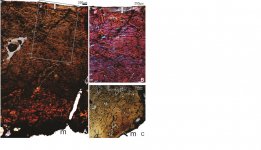
The bone microstructure of extinct animals provides a host of information about their biology. Although the giant flightless dromornithid, Genyornis newtoni, is reasonably well known from the Pleistocene of Australia (until its extinction about 50–40 Ka), aside from various aspects of its skeletal anatomy and taxonomy, not much is known about its biology. The current study investigated the histology of fifteen long bones of Genyornis (tibiotarsi, tarsometatarsi and femora) to deduce information about its growth dynamics and life history. Thin sections of the bones were prepared using standard methods, and the histology of the bones was studied under normal and polarised light microscopy. Our histological analyses showed that Genyornis took more than a single year to reach sexual maturity, and that it continued to deposit bone within the OCL for several years thereafter until skeletal maturity was attained. Thus, sexual maturity and skeletal maturity were asynchronous, with the former preceding the latter. Our results further indicated that Genyornis responded to prevailing environmental conditions, which suggests that it retained a plesiomorphic, flexible growth strategy. Additionally, our analyses of the three long bones showed that the tibiotarsus preserved the best record of growth for Genyornis.
Enjoy,
Fred
Enjoy,
Fred