

Fred Ruhe
Well-known member

Case Vincent Miller, Michael Pittman, Thomas G. Kaye, Xiaoli Wang, Jen A. Bright & Xiaoting Zheng, 2020
Disassociated rhamphotheca of fossil bird Confuciusornis informs early beak reconstruction, stress regime, and developmental patterns
Communications Biology. 3: Article number 519.
doi:10.1038/s42003-020-01252-1
Free pdf and abstract: https://www.nature.com/articles/s42003-020-01252-1.pdf
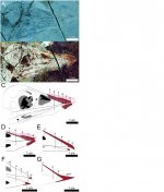
Soft tissue preservation in fossil birds provides a rare window into their anatomy, function, and development. Here, we present an exceptionally-preserved specimen of Confuciusornis which, through Laser-Stimulated Fluorescence imaging, is identified as preserving a disassociated rhamphotheca. Reconstruction of the in vivo position of the rhamphotheca validates the association of the rhamphotheca with two previous confuciusornithid specimens while calling that of a third specimen into question. The ease of dissociation is discussed and proposed with a fourth specimen alongside finite element analysis as evidence for preferential soft-food feeding. However, this proposition remains tentative until there is a better understanding of the functional role of beak attachment in living birds. Differences in postrostral extent and possibly rhamphotheca curvature between confuciusornithids and modern birds hint at developmental differences between the two. Together, this information provides a wealth of new information regarding the nature of the beak outside crown Aves.
Enjoy,
Fred
Disassociated rhamphotheca of fossil bird Confuciusornis informs early beak reconstruction, stress regime, and developmental patterns
Communications Biology. 3: Article number 519.
doi:10.1038/s42003-020-01252-1
Free pdf and abstract: https://www.nature.com/articles/s42003-020-01252-1.pdf
Soft tissue preservation in fossil birds provides a rare window into their anatomy, function, and development. Here, we present an exceptionally-preserved specimen of Confuciusornis which, through Laser-Stimulated Fluorescence imaging, is identified as preserving a disassociated rhamphotheca. Reconstruction of the in vivo position of the rhamphotheca validates the association of the rhamphotheca with two previous confuciusornithid specimens while calling that of a third specimen into question. The ease of dissociation is discussed and proposed with a fourth specimen alongside finite element analysis as evidence for preferential soft-food feeding. However, this proposition remains tentative until there is a better understanding of the functional role of beak attachment in living birds. Differences in postrostral extent and possibly rhamphotheca curvature between confuciusornithids and modern birds hint at developmental differences between the two. Together, this information provides a wealth of new information regarding the nature of the beak outside crown Aves.
Enjoy,
Fred