Systematic paleontology
Order PELECANIFORMES Sharpe, 1891
Family PELECANIDAE Rafinesque, 1815 (sensu Bock 1994).
Genus PELECANUS Linnaeus, 1758
Pelecanus sp.
Material. Distal end of a right humerus, MHN-UABCS TR27-164, collected by G. González-Barba, 9 November 1997.
Locality and age. The fossil locality is approximately 300 m northeast of Racho La Trinidad (Fig. 1), and is in biozone NN11a-b, late Tortonian-Messinian (late Miocene).
Description. The bone from the Trinidad Formation is strongly crushed and slightly twisted due to diagenetic processes, but referred to the extant Pelecanidae because of its large size, and characters that match those found in humeri of different species of Pelecanus: relatively flat in the vicinity of the wide and well-developed fossa musculi brachialis;
condylus dorsalis with a distinct hook proximally; a wide, shallow trough between the condylus dorsalis and the epicondylus dorsalis; and a distinct pit on the dorsal side between the distal end of the condylus dorsalis and the epicondylus dorsalis. There is an oblique
fracture (Fig. 3F) in the condylus ventralis, with slight displacement, and the condyle is partly worn away. In cranial view, just distad of the break of the bone shaft is a proximodistally elongate foramen.
Comparisons. The shaft of the Trinidad Formation bone appears larger than in life in both cranial and caudal views principally because of flattening. The condyles appear to have been slightly larger than P. occidentalis, more like the proportions of P. erythrorhynchos, but
with a bone probably about the same overall length as P. occidentalis (Fig. 3). The fossil has the same slight curvature of the shaft as the humerus of P. occidentalis and a foramen in almost the same position (relative to the distance from the distal end) as the same foramen in
the humerus of P. occidentalis. In overall size, the Trinidad Formation Pelecanus was much smaller than P. schreiberi Olson, 1999. The Trinidad Formation Pelecanus may have been closely related to extant P. occidentalis, with perhaps slightly more robust wing bones, but more cannot be said until more complete material is found. In a preliminary report (González-Barba et al., 2004), we had mistakenly identified the
Trinidad Formation bone as that of a pelagornithid. After further preparation of the specimen, we have now been able to better reinterpret the the crushed and distorted fossil, and directly compare the specimen with other pelagornithid specimens from western North America. The Trinidad Formation fossil is much smaller overall than the humerus of Pelagornis sp. (Fig. 4), and in Pelagornis the shaft is very straight, the fossa musculus brachialis is not as dorsoventrally wide as in Pelecanus, and the epicondylus dorsalis is prominent and flattened
in Pelagornis but rounded, not projecting in Pelecanus (see Fig. 3 for key to osteological features).
Fred
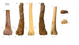
Figure 3. Right humeri of Pelecanus erythrorhynchos (A), Pelecanus sp. (MHN-UABCS TR27–164; B, D, F) and P. occidentalis (C, E, G) in cranial (A-C), caudal (D-E), and distal (F-G) views. Diagonal line in F indicates oblique fracture with flattening and displacement caudally of approximately half of the condylus ventralis. Abbreviations: cv, condylus dorsalis; cv, condylus ventralis; ed, epycondylus dorsalis; f, foramen; fmb, fossa musculus brachialis; fo, fossa olecrani; sh, sulcus humerotricipitalis; ss, sulcus scapulotricipitalis.