• Welcome to BirdForum, the internet's largest birding community with thousands of members from all over the world. The forums are dedicated to wild birds, birding, binoculars and equipment and all that goes with it.

    Please register for an account to take part in the discussions in the forum, post your pictures in the gallery and more.
ZEISS DTI thermal imaging cameras. For more discoveries at night, and during the day.

12 new birds from the Miocene of Argentina (1 Viewer)

Fred Ruhe

Well-known member
Netherlands
A paper I overlooked:

Federico Lisandro Agnolin, 2022

New fossil birds from the miocene of Patagonia, Argentina
Nuevas aves fósiles del mioceno de Patagonia, Argentina


Poeyana Revista Cubana ddr Zoolo9gía 513 (Januari-December 2022)

Abstract and free pdf: Vista de Nuevas aves fósiles del mioceno de Patagonia, Argentina

The present contribution aims to describe new fossil bird remains coming from early-middle Miocene beds (Pinturas and Santa Cruz Formations) in Santa Cruz province, Patagonia, Argentina. These include a new tinamid, a basal anseriform, one anhimid, two new tadornine ducks, possible gruid, psophiid, and parvigruid cranes, an uncertain falconid, a new species of the falconid genus Thegornis, a possible sophiornithid, a coraciid, and remains of innominate rallids, phoenicopterids and passeriforms of the clade Tyranni. If correctly identified, the gruid represents the oldest record for the clade in South America and one of the few findings of the group in the entire continent. The sophiornithid and parvigruid may constitute the first record for each clade in South America. The psophiid may constitute the first fossil record for the clade worldwide. The coraciid represents the first record for the family in South America and both the youngest and second record for coraciiforms in the continent. This, together with the gruid, constitute members of bird clades that were geographically widespread by Paleogene and early Neogene times, but now are restricted as relicts to the Old Word and North America. Their extinction from the Neotropical Region is still uncertain. The presence of at least three different members of Tyranni, reinforces previous thoughts sustaining a long and complex history of the subclade, and passeriforms as a whole, in South America.

Enjoy,

Fred
 
SYSTEMATIC PALEONTOLOGY

Palaeognathae Pycraft, 1900
Tinamidae Huxley, 1872
Genus Mininothura nov. gen.

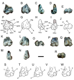
Diagnosis. Very small tinamid, smaller than members of the genus Nothura and diagnosable on the basis of the following unique combination of characters: 1- relatively wide and well-defined brachial fossa; 2- anterior articular ligament not raised and subcircular in contour; 3- ectepicondylar process not raised, forming a prominent and thickened lateral edge; and 4- ventral condyle distally extended and subequal in size to dorsal condyle.

Etymology. Mini, meaning small in Latin, Nothura, a tinamid genus.

Type and only known species. Mininothura talenki nov. sp.

Mininothura talenki nov. sp.

Diagnosis. The same as for genus by monotypy.

Holotype. MACN SC 1429, incomplete distal end of left humerus

Paratypes. MACN SC 1612, incomplete distal end of right humerus; MACN SC 1427, incomplete distal end of left humerus

Locality and horizon. Loma de la Lluvia fossil site, Pinturas river, Santa Cruz province, Argentina. The specimens come from beds belonging to the Early- Middle Miocene Pinturas Formation

Etymology. From the Aonikenk language, talen meaning small.

Remarks. Mininothura shows a combination of characters present in tinamids, including the distal end of the humerus being transversely wide and anteroposteriorly flat, dorsal and ventral condyles subequal in size, poorly excavated brachial fossa, a wide and ovoidal-shaped flat surface proximal to the dorsal condyle, a wide, flattened, and subcircularshaped surface separating the ventral condyle from the dorsal condyle and from the facet of the anterior articular ligament (see Bertelli, 2002, 2017; Bertelli and Chiappe, 2005; Bertelli et al., 2014). Extant tinamids are distributed in two main clades: Nothurinae or open-area tinamous, and Tinaminae or forest-dweeling tinamous (Bertelli, 2017; Bertelli and Chiappe, 2005; Bertelli et al. ,2014)
Mininothura shares with tinamines a rounded ectepicondylar process, a character that is synapomorphic of the clade (Bertelli and Chiappe, 2005). However, Mininothura is unique in the shape of the ectepicondylar process, which is represented by a poorly defined intumescense that is continuous with the lateral margin of the bone. Whether this condition is homologous to that of tinamines is far from certain, and thus, Mininothura is not assigned to any particular tinamid subclade.
The small size of the material may fall within the range of the genera Nothura, Crypturellus, Nothoprocta or Taoniscus, being much smaller than other tinamids (Cenizo et al. , 2012; Chandler, 2012). Comparisons with other tinamids reinforces the
distinctiveness of Mininothura. In Mininothura the brachial fossa is obliquely oriented with respect to the axis of the bone shaft and is oval in contour, a morphology shared with Tinamus, Eudromia, Tinamotis, Rhynchotus and Nothocercus, and different from the crescent-shape observed in Nothura, Nothoprocta and Peioa (Bertelli et al. , 2014). The brachial fossa is more excavated than in other small tinamids, such as Nothura and Nothoprocta, resembling Toaniscus in this aspect (Bertelli et al., 2014). The flexor process is relatively short, not extended beyond the ventral condyle, and very different from the elongate condition present in Nothocercus, Taoniscus, Nothoprocta and Rhynchotus (Bertelli et al., 2014). Mininothura lacks the secondary process close to the supracondylar dorsal process observed in Tinamus and some Crypturellus species (Bertelli, 2002). The ventral condyle is proximally undercut, whereas in Tinamus, Nothocercus, Crypturellus and Taoniscus it is poorly delimited proximally (Cenizo et al. , 2012). The dorsal condyle is subequal in size to the ventral one, a
condition shared with Tinamotis (Bertelli and Chiappe, 2005). The facet for the anterior articular ligament forms a shallow and well-differentiated subcircular concavity that differs from the less defined and shallower condition observed in
Tinamus, Nothocercus and Crypturellus (Cenizo et al., 2012).
Bertelli and Chiappe (2005) reported the distal end of tinamid humeri as coming from early-middle Miocene beds of the Santa Cruz Formation, in Santa Cruz province. The two specimens described by Bertelli and Chiappe (2005) belong to two closely related, but different species. These specimens can be clearly distinguished from Mininothura by having ventral condyle notably larger than the dorsal one, by the much smaller and shallow brachial fossa, and by a prominent ectepicondylar process. On this basis, it can be affirmed that at least three different smallsized and one large tinamid were present in Santa Cruz province during early-middle Miocene times.

Fred


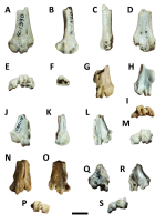
Figure 1. Mininothura talenki A-J, incomplete distal end of left humerus (MACN SC 1429, holotype) in A,F, anterior; B,G, posterior; C,H, medial; D,I, lateral; and E,J, distal views; K-O, incomplete distal end of right humerus (MACN SC 1612, paratype) in K, anterior; L, posterior; M, lateral; N, medial; and O, lateral views; P-R, incomplete distal end of left humerus (MACN SC 1427, paratype) in P, anterior; Q, posterior; and R, distal views. S-W, distal end of left humerus of selected tinamids in anterior view; S, Crypturellus tataupa; T, Nothoprocta cinerascens; U, Tinamotis pentlandii; V,
Figure 2. Mininothura talenki A-J, incomplete distal end of left humerus (MACN SC 1429, holotype) in A,F, anterior; B,G, posterior; C,H, medial; D,I, lateral; and E,J, distal views; K-O, incomplete distal end of right humerus (MACN SC 1612, paratype) in K, anterior; L, posterior; M, lateral; N, medial; and O, lateral views; P-R, incomplete distal end of left humerus (MACN SC 1427, paratype) in P, anterior; Q, posterior; and R, distal views. S-W, distal end of left humerus of selected tinamids in anterior view; S, Crypturellus tataupa; T, Nothoprocta cinerascens; U, Tinamotis pentlandii; V, Mininothura talenki; and W, Eudromia elegans. Abbreviations. aal, attachment for the anterior articular ligament; bf, brachial fossa; dc, dorsal condyle; ect, ectepicondylar process; en, entepicondylar process; ft, flexor tubercle; htg, humerotricipital groove; mcu, attachment of the m. carpi ulnaris; meu, surface for the m. ectepicondyloulnaris; mps, pit for the m. pronator superficialis; of, olecranal fossa; sct, scapulotricipital groove; spdc, flat surface proximal to the dorsal condyle; spvc, flat surface proximal to the ventral condyle; su, surface for the m. supinator; vc, ventral condyle. Remarkable diagnostic features of selected tinamids: 1, small and crescent-shaped brachial fossa; 2, well-developed and prominent ectepicondylar process; 3, small and rounded ectepicondylar process; 4, distally extended and prominent flexor process. Scale bar: A-R, 5 mm; S-W, not to scale.
1694411825522.png
 
Last edited:
Anseriformes Wagler, 1831
Genus Peioa nov. gen.

Diagnosis. Medium-sized anseriform, having the following unique combination of characters: 1- brachial fossa crescent-shaped, well-defined, and notably narrow; 2- brachial fossa obliquely oriented, with its distal third subvertically oriented, very close
and subparallel to the medial edge of the shaft; 3- surface for the anterior articular ligament anteriorly protrudent and distally facing; 4- ectepicondylar process prominent, but located distal to the proximal level of the dorsal condyle; and 5- humerotricipital groove well-defined and delimited by relatively sharp ridges.

Etymology. Peio, from the Aonikenk language, meaning hen.

Type and only known species. Peioa australis nov. sp.

Peioa australis nov. sp.

Diagnosis. The same as for genus by monotypy.

Holotype. MACN SC 1452, distal half of right humerus

Locality and horizon. The specimen comes from the Santa Cruz Formation (Early-Middle Miocene), at the well-known fossiliferous locality of Monte Observación (the label indicates “Estaca 29”), Monte León National Park, Santa Cruz province, Argentina.

Etymology. Australis, from the Latin language, meaning southern.

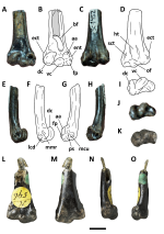
Remarks. Peioa australis nov. sp. shows a combination of characters that indicate anseriform affinities: a low ectepicondylar process, a flattened entepicondyle with subparallel, well-defined, and oval pits for the m. carpi ulnaris and m. pronator superficial, and an ectepicondylar process with subparallel, well-defined, and oval pits for the m. carpi radialis and lig. collateral fossa (Worthy et al.,2022). In spite of being clearly anseriform, the morphology of Peioa is very different from most ducks and geese.
The distal end of the humerus in Peioa nov. gen. is remarkable in being transversely expanded, anteroposteriorly compressed, with a very small and poorly excavated musculus brachialis antiquus impression, and the shaft poorly differentiated from the distal end of the bone (Woolfenden 1961, Olson 1999; Cenizo and Agnolín, 2010), similar to the condition observed in the basal anseriforms Conflicto, Anseranas and Anatalavis (Olson 1999; Tambussi et al. , 2019; Worthy et al. , 2022). However,
Peioa clearly differs from the later taxa in some anatomical details. In Peioa the surface for the anterior articular ligament is anteriorly protrudent and distally facing, it is transversely wide and proximodistally low. In contrast, in Conflicto, Anatalavis and Anseranas, this surface is not prominent and ovoidal in contour, being more proximodistally tall than transversely wide. The flexor process in Peioa is much more distally extended than in Anatalavis and Anseranas, and the brachial fossa is smaller. In Peioa the ectepicondylar process is prominent, but is located distal to the proximal level of the dorsal condyle, whereas in Anatalavis and Anseranas it is located more
proximally (Olson, 1999). In contrast to Anseranas and reminiscent to Anatalavis, in Peioa the olecranal and humerotricipital fossae are deep and welldefined (Olson, 1999). The shape of the brachial fossa in Peioa clearly differs from that of Anatalavis
and Anseranas.
Eutelornis patagonicus was described by Ameghino (1894) based on associated distal radius, distal humerus and partial tibiotarsus from the Santa Cruz Formation, in Santa Cruz province, Argentina. Ameghino regarded Eutelornis as an anseriform of uncertain affinities. Later, because of its plesiomorphic-looking aspect, Cenizo and Agnolín (2010) indicated that it was a very basal anseriform of uncertain affinities, probably related to Anseranatidae.
Eutelornis is somewhat reminiscent to Peioa in having the distal end notably flattened, the brachial fossa not strongly excavated and crescent-shaped, and its distal end subvertically oriented and subparallel to the medial margin of the humeral
shaft. Furthermore, it shows a very lare ventral condyle, a condition similar to that of
palaeognathous tinamids (Bertelli and Chiappe, 2005). Peioa differs from Eutelornis in having a smaller ventral condyle, in having a rounded and not prominent supracondylar process that is more distally positioned, and in having well-defined humerotricipital groove and ridges.
Based on the presence of a large number of common morphological attributes, it is probable that Peioa and Eutelornis may form a clade of basal anseriforms.

Fred


Figure 1. A-J, Peioa australis, distal half of right humerus (MACN SC 1452, holotype) in A-B, anterior; C-D, posterior; E-F, medial; G-H, lateral; and I-J, distal views. K-O, Eutelornis patagonicus, distal half of right humerus (BMNH A-596, holotype) in K, distal; L, anterior; M, posterior; N, medial; and O, lateral views.
Abbreviations. aa, surface for the anterior articular ligament; bf, brachial fossa; ect, ectepicondylar process; ent, entepicondylar process; fp, flexor process; ht, humerotricipital groove; lcd, scar of the dorsal collateral ligament; mcu, scar for the m. carpi ulnaris; mmr, scar for the m. metacarpi radialis; of, olecranon fossa; ps, scar for the pronator superficial; sct, scapulotricipital groove. Eroded areas shaded in grey. Scale bar 1 cm. Photographs K-O, courtesy of Sandra Chapman (BMNH) and Jorge Noriega. Scale bar 5 mm.
1694415577857.png
 
Last edited:
Anhimidae Stejneger, 1885
Genus Chainkanas nov. gen.

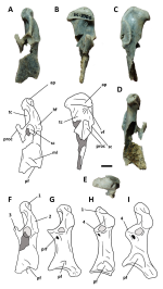
Diagnosis. Large anseriform diagnosed on the basis of the following combination of characters: 1- procoracoid extensive and dorsally hooked (shorter and not dorsally tilted in Chauna, Anhima and Chaunoides); 2- humeral facet strongly medially tilted (not tilted in Chauna, Anhima and Chaunoides); 3- humeral facet nearly reaching the level of the distal end of the scapular facet (more proximally placed in Chauna, Anhima and Chaunoides); 4- scapular facet subcircular in contour and deeply excavated, and slightly distally oriented (strongly laterally oriented in Chauna, Chaunoides and Anhima); and 5- acrocoracoid process elongated (shared with Chaunoides; much wider and stouter in Chauna and Anhima).

Etymology. Chaink, meaning large in Aonikenk language; anas, meaning duck in Latin.

Type and only known species. Chainkanas koshon nov. sp.

Chainkanas koshon nov. sp.

Diagnosis. The same as for genus by monotypy.

Holotype. MACN SC 3306, right coracoid with incomplete distal end.

Locality and horizon. Loma de la Lluvia fossil site, Pinturas river, Santa Cruz province, Argentina. The specimen comes from beds belonging to the Early-Middle Miocene Pinturas Formation.

Etymology. Koshon, from the Aonikenk language meaning scream; members of the family Anhimidae are popularly known as “screamers” in English language.

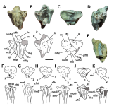
Remarks. Chainkanas nov. gen. can be included among anhimids by having very large size, robust coracoid with a large procoracoid process, procoracoid foramen extensive and ellipsoidal in contour, and the presence of a large pneumatic foramen at its distal end (Alvarenga, 1999). As indicated in the diagnosis, Chainkanas can be clearly distinguished by other extant and extinct anhimids, especially by its procoracoid shape. Among anhimids, it is more similar to the Oligocene genus Chaunoides from Brazil than to extant genera Anhima and Chauna, by having a relatively elongate coracoid, narrow acrocoracoid process, and very wide and deep scapular fossa (Alvarenga, 1999).
Loxornis clivus is a bird species based on a distal tibiotarsus with eroded distal condyles coming from the Deseadan beds (Oligocene) of Santa Cruz province. The only known specimen. It was originally described by Ameghino (1895) as an uncertain
anseriform (Tonni, 1980), and more recently it was regarded as a possible anhimid by Alvarenga (1999; see Cenizo and Agnolín, 2010). Because Loxornis and Chainkanas are based on different materials, a direct comparison between them is not possible. However, it should be pointed out, that both come from distant stratigraphical units that show strong faunal differences. Furthermore, as pointed out by Alvarenga (1999) the phylogenetic position of Loxornis is not clear, and it is not entirely certain that it belongs to Anhimidae.

Fred


Figure 1. Chainkanas koshon (MACN SC 3306, holotype), right coracoid lacking distal end in A, dorsal; B, medial; C, lateral; D, ventral; and E, proximal views. F-I, coracoid of selected anhimids in dorsal view: F, Chainkanas koshon; G, Anhima cornuta; H, Chaunoides antiquus; I, Chauna torquata. Abbreviations. ap, acrocoracoid process; hf, humeral facet; ml, muscular line; pf, pneumatic foramen; prf, procoracoid foramen;
proc, procoracoid process; sc, scapular facet; tc, triosseal canal; vf, ventral fossa. 1, relatively narrow acrocoracoid; 2, medially tilted humeral facet; 3, hooked procoracoid process; 4, thickened and short procoracoid process. Scale bar: A-E, 1 cm; F-I, not to scale. G-I, redrawn from Alvarenga (1999) and personal inspection of specimens.
1694418288639.png
 
Anatidae Leach, 1820
Tadorninae Reichenbach, 1850
Genus Kaikenia nov. gen.

Diagnosis. Relatively large anatid, very similar in size and shape to Miotadorna, having the following unique combination of characters: 1- lateral margin of the shaft nearly straight; 2- attachment of the pronator muscle distally located with respect to the proximal margin of the anterior articular ligamental facet (at the same level in remaining tadornines; Worthy et al. , 2009); 3- scapulotricipital groove deep, but does not extend around the distal end of the bone (in contrast to remaining tadornines; Worthy et al., 2009); 4- flexor process poorly extended distally; and 5- olecranal fossa in distal view, laterally delimited by a thick ridge.

Etymology. Kaiken, meaning members of the goose genus Chloephaga in Aonikenk language.

Type and only included species. Kaikenia mourerchauvirea nov. sp.

Kaikenia mourerchauvirea nov. sp.

Diagnosis. The same as for genus by monotypy.

Holotype. MACN SC 4506, right humerus lacking its proximal third.

Locality and horizon. The specimen comes from the Santa Cruz Formation (Early-Middle Miocene), at the well-known fossiliferous locality of Monte Observación (the label of the specimen indicates “Estaca 7”), Monte León National Park, Santa Cruz province, Argentina. Collected in 1991.

Referred material. MACN SC 3431, proximal end of left carpometacarpus.

Locality and horizon. The referred specimen comes from the Santa Cruz Formation (Early-Middle Miocene), Monte Observación locality, Monte León National Park, Santa Cruz province, Argentina.

Etymology. The specific epithet honors Cécile Mourer-Chauviré, a leading paleontologist from France, who greatly contributed to our current knowledge on palaeornithology.

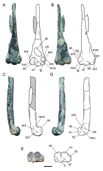
Remarks. Kaikenia nov. gen. may be referred to Tadorninae on the basis of the following combination of characteristics: tendence to narrowing of the humeral shaft, distinct ectepicondylar prominence and scapulotricipital groove, distal margin of flexor process level with distal margin of dorsal condyle, and shallow brachial fossa (Worthy, 2009; Worthy et al., 2007).
Its large size easily distinguishes Kaikenia from several small tadornines, such as Nannonetta, Pleistoanser and Anabernicula. The North America Pleistocene genus Brantadorna lacks overlaping material with Kaikenia, and thus, comparisons are
not possible (Howard, 1963).
Kaikenia differs from members of the genera Chloephaga, Neochen, Anabernicula,
Australotadorna, Pleistoanser and Alopochen in having a straight shafted and narrow humerus, with poorly developed flexor tubercle, and much less prominent ectepicondylar prominence (see Agnolín, 2006a; Howard, 1964; Worthy, 2009). In these features, Kaikenia resembles tadorninines of the genera Tadorna and Miotadorna. However, it differs from both by having a prominent entepicondylar process and the attachment for the m. carpi ulnaris very deep and medially oriented (Worthy et al. , 2007). Particularly, Kaikenia is similar in shape and development of the ectepicondylar process, flexor process, and facet for the anterior articular attachment to the extinct Miocene genus Miotadorna (Worthy and Lee, 2008; Worthy et al., 2007). However, it differs in several important anatomical features (as indicated above), including a shallower brachial fossa (distally very deep in Miotadorna, Alopochen and Australotadorna; Worthy, 2009) and having a poorly distally oriented flexor process (Worthy et al., 2007). A feature that occurs in Kaikenia, that is unknown in other
tardornines, is the presence in distal view of a ridge that delimits the olecranal fossa. This feature may be regarded as autapomorphic of Kaikenia.
Teleornis impressus is an anatid named by Ameghino 1899 on the basis of an incomplete forelimb coming from Deseadan Oligocene beds at Santa Cruz province. Teleornis was later considered as belonging to Tadorninae (Agnolín, 2004). Kaikenia differs from Teleornis in being much larger and in having the distal end of the bone transversely wider and more anteroposteriorly flattened.
Furthermore, Kaikenia differs in having a much thicker and less distally extended flexor process, and by the shallower humerotricipital groove.

Fred


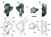
Figure 1. Kaikenia mourerchauvirea A-E, right humerus lacking proximal end (MACN SC 4506, holotype) in A, anterior; B, posterior; C, lateral; D, medial; and E, distal views. Abbreviations. aal, surface for the anterior articular ligament; bf, brachial fossa; ect, ectepicondylar process; ent, entepicondylar process; fp, flexor process; hg, humerotricipital groove; lcd, scar of the dorsal collateral ligament; lr, lateral ridge; mcu, scar for the m. carpi ulnaris; mmr, scar for the m. metacarpi radialis; of, olecranon fossa; ps, scar for the pronator superficial; sg, scapulotricipital groove. Scale bar 1 cm.
1694420595861.png

Figure 2. Kaikenia mourerchauvirea A-D, proximal end of left carpometacarpus (MACN SC 3431, referred material), A, medial; B, lateral; C, proximal; and D, posterior views. Eroded areas shaded in grey. Abbreviations. afc, anterior carpal fossa; ap, alular process; dei, distal extension of medial rim of carpal trochlea; ep, extensor process; etr, external carpal trochlea; if, infratrochlear fossa; ma, major metacarpal; mi, minor metacarpal; pcf, posterior carpal fossa; pis, pisiform process; sf, supratrochlear fossa; sy, proximal synostosis. Scale bar: 1 cm.
1694420715935.png
 
Genus Tamtamia nov. gen.

Diagnosis. Mid-sized and relatively gracile tadornine diagnosable on the basis of the following combination of characteristics: distal end of humerus: 1- very low ectepicondylar prominence that is distally located level with the dorsal margin of the dorsal condyle; 2- very prominent and large attachment for the anterior articular ligament, which is very prominent and medially displaced; 3- attachment for the anterior articular ligament ventrally delimited by a strongly concave fossa for the m. carpi ulnaris; 4- small flexor process that is not distally extended and is located close to the dorsal margin of the ventral condyle; proximal end of humerus: 5- transversely expanded and anteroposteriorly flattened; 6- shallow pneumotricipital fossa; 7- ventral pneumotricipital fossa pneumatic, and delimited by lateral and wellraised ridge (dorsal crus commune); 8- bicipital crest extensive and strongly flared; and 9- capital ridge located between the humeral head and the dorsal tubercle.

Etymology. Támtam, meaning female members of the goose genus Chloephaga in Aonikenk language.

Type and only included species. Tamtamia yzurietai nov. sp.

Tamtamia yzurietai nov. sp.

Diagnosis. The same as for genus by monotypy.

Holotype. MACN SC 4507, distal end of right humerus and proximal end of left humerus from the same individual.

Paratypes.
MACN SC, 4508, distal end of left humerus; MACN SC 3309, proximal end of right humerus.

Locality and horizon. Loma de la Lluvia fossil site, Pinturas river, Santa Cruz province, Argentina. The specimen comes from beds belonging to the Early- Middle Miocene Pinturas Formation.

Etymology. Yzurietai, honouring the naturalist, birdwatcher and artist Dario Yzurieta (1931-1996). Together with “Tito” Narosky they published the most useful Bird Guide Identification of the Neotropics. Due to their conjoined efforts, hundreds of young people become birdwatchers (including the author of the present contribution).

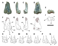
Remarks. The distal end of the humerus of Tamtamia nov. gen. is very different from basal forms such as anseranatids, anhimids, dendrocygnines and romainvilliines by being anteroposteriorly thicker and transversely narrow, and by the poorly excavated distal end of the bone proximal to the ventral and dorsal condyles (Agnolín and Tomassini, 2012; Mayr, 2008).
Tamtamia is assigned to Tadorninae on the basis of the following combination of characteristics: prominent capital shaft ridge located close to the level of the dorsal tubercle, elevated and elongate dorsal tubercle, dorsal pneumotricipital fossa narrow and not strongly excavated under the humeral head, large ventral pneumotricipital fossa, deltoid crest concave in posterior view, and strongly raised and distally oriented facet for the anterior articular ligament (Woolfenden, 1961; Worthy, 2009).
Tamtamia differs from Chloephaga in that it lacks the very weak capital ridge and the shaft compression adjacent to the angle of the deltoid crest, but well rounded from there, a condition unique to this genus (Worthy, 2009). It also differs in the more proximodistally narrow and less prominent humeral head. It differs from Pleistocene
Anabernicula, Pleistoanser and Nannonetta in the poorly distally extended flexor process and in the distally positioned ectepicondylar prominence, among several other anatomical details (Agnolín, 2006a; Howard, 1964). From Neochen, Alopochen and Brantadorna it differs in the much more gracile proportions, narrower shaft, and smaller and less prominent humeral head (see Howard, 1963; Short, 1970). It differs from Australotadorna in having a more gracile construction, with a less prominent humeral head, ventral pneumotricipital fossa delimited by a ridge, and rounded and relatively low ventral tubercle, among several other anatomical traits (see Worthy, 2009).
In size and proportions, Tamtamia is reminiscent to the extant genus Tadorna and extinct Miocene genus Miotadorna (see Worthy and Lee, 2008; Worthy et al., 2007). It differs from Tadorna in having a much wider and excavated ventral pneumotricipital fossa that is laterally delimited by a subvertical ridge, by having a more prominent but distally located ectepicondylar prominence, and a proximally positioned flexor process. It differs from Miotadorna in having a notably wider and more excavated ventral pneumotricipital fossa, and a prominent and distally located ectepicondylar prominence (Worthy and Lee, 2008; Worthy et al., 2007).
Tamtamia differs from Kaikenia (described above) in several features, including a deeper and more extensive humerotricipital groove, a well-raised facet for the anterior articular ligament, a deep brachial fossa, and different development of the
ectepicondylar process and flexor process. Tamtamia differs from Teleornis (Ameghino, 1899; Agnolín, 2004) in that the latter has a prominent and distally extended flexor process, whereas in Tamtamia it is much shorter and more proximally positioned with respect to the distal end of the ventral condyle. The ventral condyle in Tamtamia is subhorizontally oriented, whereas in Teleornis it is strongly oblique,
forming an angle of about 90° with the main axis of the dorsal condyle (see Agnolín, 2004).
Tamtamia comes from beds that are probably coeval with those that yielded the basal anatid Ankonetta larriestrai (Cenizo and Agnolín, 2010). Regrettably, Ankonetta is based on an incomplete tarsometatarsus, and thus, there is no overlapping material with Tamtamia. As described by Cenizo and Agnolín (2010), Ankonetta is a basal dendrocygnine like anatid, and in all probability larger in size than that inferred for Tamtamia. It is not improbable that after the finding of new materials, Tamtamia may
result in being a junior synonym for Ankonetta .

Fred


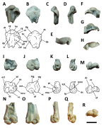
Figure 1. Tamtamia yzurietai. A-E, I-M, distal end of right humerus and proximal end of left humerus (MACN SC 4507, holotype) in A, J, posterior; B,I, anterior; C,K, medial; D,L, lateral; and E,M, distal views. F-H, proximal end of right humerus (MACN SC 3309, paratype) in F, posterior; G, anterior; and H, proximal views. NR, distal end of left humerus (MACN SC, 4508, paratype) in N, anterior; O, posterior; P, lateral; Q, medial; and R, distal views. Abbreviations. aa, surface for the anterior articular ligament; bc, bicipital crest; bf, brachial fossa; cdf, crus dorsale fossae; cg, capital groove; cr, capital ridge; cvf, crus ventrale fossae; dt, dorsal tubercle; dp, deltopectoral crest; ect, ectepycondyle; ent, entepicondylar process; fp, flexor process; he, humeral head; htg,
humerotricipital groove; int, intumescencia; lcd, scar of the dorsal collateral ligament; mcu, scar for the m. carpiulnaris; mmr, scar for the m. metacarpi radialis; of, olecranon fossa; pne, pneumotricipital fossa; ps, scar for the pronator superficial; stg, scapulotricipital groove; vt, ventral tubercle. Scale bar 1 cm.
1694422435001.png
 
Phoenicopteriformes Sharpe, 1891
Indeterminate genus and species

Referred material. MACN SC 1401, distal end of left humerus (Figure 8A-E); MACN SC 4509, distal end of right tarsometatarsus lacking II metatarsal trochlea

Locality and horizon.
Loma de la Lluvia fossil site, Pinturas river, Santa Cruz province, Argentina. The specimen comes from beds belonging to the Early-Middle Miocene Pinturas Formation.

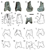
Remarks. The specimens described here can be referred to Phoenicopteriformes on the basis of the following combination of characteristics: high and flattened ventral supracondylar tubercle, flexor process not posteriorly extended, well-defined olecranal fossa, tarsometatarsus strongly transversely compressed, trochlea IV with very wide and expanded posterior wing, and proximally positioned vascular foramen (Ericson, 1999; Feduccia, 1976; Zelenkov, 2021).
Among phoenicopteriforms, MACN SC 1401 and MACN SC 4509 are reminiscent to stemphoenicopteriforms, such as Juncitarsus, in being relatively small in size, having strong distal compression of the tarsometatarsus, and in having a small and obliquely oriented ectepicondylar process on the humerus (Ericson, 1999). These features distinguish them from modern phonicopteriforms, such as Phoenicopterus, Phoeniconaias and Palaelodus (Vickers Rich et al. , 1987). In this way, we regard these specimens as probably being related to stem-phoenicopteriforms like Juncitarsus.

Fred


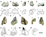
Figure 1. Indeterminate Phoenicopteriformes. A-E, distal end of left humerus (MACN SC 1401) in A, anterior; B, posterior; C, lateral; D, medial; and E, distal views; F-N, distal end of right tarsometatarsus lacking II metatarsal trochlea (MACN SC 4509) in F, anterior; G, lateral; H, medial; I, distal; and J, posterior views. Abbreviations. aa, anterior articular ligament; dc, dorsal condyle; ect, ectepicondylar process; ent, entepicondylar process; fp, flexor process; htg, humerotricipital groove; lcd, scar of the dorsal collateral ligament; mccol, surface for attachment of the m. carpi radialis and the collateral ligament; mcu, scar for the m. carpi ulnaris; of, olecranal fossa; plw, posterolateral wing; scg, scapulotricipital groove; vc, ventral condyle. II-IV, distal metatarsal trochleae. Eroded areas shaded in grey. Scale bar: 1 cm.
1694425707941.png
 
Gruiformes (Bonaparte, 1854)
Rallidae Vigors, 1825
Indeterminate genus and species

Referred material. MACN SC 1431, incomplete d tal end of right tarsometatarsus MACN SC 1433, proximal end of left tarsometatarsus.

Locality and horizon. Loma de la Lluvia fossil site, Pinturas river, Santa Cruz province, Argentina. The specimen comes from beds belonging to the Early-Middle Miocene Pinturas Formation.

Remarks. The specimens reported here can be referred to Rallidae on the basis of a unique combination of characteristics, including a hypotarsus with a prominent lateral crest and a reduced medial one, intercotylar eminence relatively low and well-defined, and anteriorly projected, distal end of the bone with proximally extended articular surfaces of trochleae, trochlea II proximally positioned and tilted, and a large and prominent scar proximal to metatarsal III in posterior view (Mayr, 2006, 2016a, 2019; Mayr and Smith, 2001). Among rallids, the position of the specimens reported here is uncertain. The size and proportions are very similar to members of the genus Rallus (Olson, 1974). They lack the flattened tarsometatarsus with an acute medial edge diagnostic of Porphyrio and kin (Mayr, 2019; Olson, 1973). The metatarsal II trochlea is proximally positioned, in contrast with the more distal location observed in Himantornis (Olson, 1973).
Regrettably, the incomplete nature of the specimens reported here precludes a more accurate phylogenetic position among rallids.

Fred


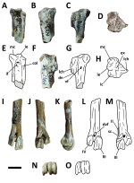
Figure 1. Indeterminate Rallidae. A-H, proximal end of left tarsometatarsus (MACN SC 1433) in A,E, anterior; B, posterior; C,G, medial; D,H, proximal; and F, lateral views. I-O, incomplete distal end of right tarsometatarsus (MACN SC 1431) in I,L, anterior; J,M, posterior; K, lateral; and N-O, distal views. Abbreviations. col, surface for the colateral lateral ligament; dn, distal notch; dvf, distal vascular foramen; ex, proximal excavation; ie, intercotylar eminence; if, infracotylar fossa; lc, lateral cotye; lch, lateral crest of hypotarsus; mc, medial cotyle; sc, muscular scar; vf, proximal vascular foramen; II, base of metatarsal trochlea II; III-IV, distal metatarsal trochleae. Eroded areas shaded in grey. Scale bar 5 mm.
1694426262942.png
 
Gruoidea sensu Wetmore, 1960
Gruidae Vigors, 1825
?Gruinae Vigors, 1825
Genus Patagogrus nov. gen.

Diagnosis. Medium-sized gruid diagnosed on the basis of the following combination of characters: 1- distal end of the bone with its anteroposterior length subequal to transverse width (shared with Palaeogrus; much transversely wider than anteroposteriorly long in Grus); 2- intercondylar groove in distal view narrow and with subparallel lateral and medial margins (relatively wide and forming an opened “V” or “U” in Grus and Palaeogrus ); 3- distal end of lateral condyle with a well-developed notch (shared with Grus, nearly absent in Palaeogrus); 4- medial condyle strongly anteriorly projected and with its anterior end acute and transversely narrow (shared with Palaeogrus; narrower in Grus); 5- medial epicondyle poorly developed (well-developed and exposed when viewed distally in Grus and Palaeogrus); 6- tubercle lateral to supratendinal bridge poorly developed (shared with Palaeogrus, in Grus the tubercle is very strong); and 7- proximodistally extended supratendinal bridge (shared with Grus).

Etymology. Patago, from Patagonia, and Grus , the type genus of Gruidae.

Type and only included species. Patagogrus olsoni nov. sp.

Patagogrus olsoni nov. sp.

Diagnosis. The same as for genus by monotypy.

Holotype. MACN SC 1423, distal end of right tibiotarsus

Locality and horizon. Loma de la Lluvia fossil site, Pinturas river, Santa Cruz province, Argentina. The specimen comes from beds belonging to the Early-Middle Miocene Pinturas Formation.

Etymology. The specific epithet honours Storrs L. Olson (1944-2020), one of the most important paleornithologists from the XX and XXI centuries.

Remarks.
Patagogrus nov.gen. shows a unique combination of characteristics shared with Gruidae, including flat anterior face of the tibiotarsus, proximodistally low distal trochlea in posterior view, anteriorly divergent condyles, lateral condyle anteroposteriorly long, with a shallow notch on its distal margin, medial condyle more anteriorly projected than the lateral one, anterior end of medial condyle transversely narrow, the presence of a tubercle lateral to the base of the supratendinal bridge, well developed ridge delimiting the lateral margin of the extensor groove, and supratendinal bridge proximodistally tall (Clarke et al., 2005;
Cracraft, 1973a; Livezey, 1998; Mayr, 2013a).
Patagogrus differs from psilopterine phorusrhacids by having a slimmer configuration, more dorsoventrally lower and transversely wider distal condyles, by having a subelliptical external rim on the lateral condyle, a medial condyle much more deflected medially, not alligned with the medial margin of the shaft, an anterior intercondylar fossa, and a poorly excavated extensor groove, among other anatomical details (see Noriega et al. 2009).
Patagogrus clearly differs from cariamids in having the lateral condyle not anteriorly projected and with a reduced epicondyle, a proximodistally high supratendinal bridge, a transversely wide and proximodistally low distal opening of the
supratendinal bridge, the presence of a tubercle lateral to the supratendinal bridge, and absence of a ridge connecting the lateral edge of the supratendinal bridge with the medial margin of the lateral condyle, among other features (Cracraft, 1968; Noriega et al., 2009).
Among gruids, Patagogrus clearly differs from Balearica and extinct Balearicinae, previously referred to as Probalearica (Mourer Chauviré, 2001), in that balearicines exhibit a robust distal end of the tibiotarsus with a strongly anteroposteriorly flattened shaft, a strongly medially deflected medial condyle, and a transversely expanded extensor groove (Feduccia and Voorhies, 1992; Mourer Chauviré, 2001). On the other side, Patagogrus resembles gruines in having a progressive (not abrupt) widening of the distal end of the bone, in the presence of a strong tubercle lateral to the supratendinal bridge, a relatively long lateral condyle, a transversely narrow extensor groove occupying only the medial part of the cranial face of the bone, and a notch at the distal edge of lateral pronounced in the Balearicinae (Göhlich, 2003; Mourer Chauviré, 2001).
Tertiary gruines are still poorly known. Several previously published records rest on weak evidence and they are considered to be of uncertain phylogenetic affinities (Cracraft, 1973a; Göhlich, 2003), which makes comparisons of Patagogrus with other fossil gruines difficult. The only Tertiary extinct gruine genera that rest on relatively robust evidence are Palaeogrus and Camusia (e.g., Göhlich, 2003; Mayr et al., 2020b; Mourer-Chauviré, 2001; Seguí, 2002; Zelenkov, 2015). Comparisons between Patagogrus, Palaeogrus and the extant genus Grus were made in the diagnosis of Patagogrus following characteristics noted by previous authors (Cracraft, 1973a; Göhlich, 2003; Northcote and Mourer-Chauviré, 1988). Camusia is very different from
Patagogrus and any other gruid genus (Seguí, 2002). It differs from Patagogrus in several anatomical details, including a tibiotarsus with a much transversely wider and anteroposteriorly flattened distal end, by having narrower and a more medially projected medial condyle and a prominent and elongate medial epicondyle.
The combination of characteristics enumerated above indicates that Patagogrus is a valid gruid genus. Among other anatomical details, it is distinctive in having a transversely narrow distal end of the tibiotarsus, as is shown by the shape of the intercondylar groove in distal view.

Fred


Figure 1. Patagogrus olsoni A-H, K, distal end of right tibiotarsus (MACN SC 1423) in A,E, anterior; B,F, posterior; C, lateral; D,H, medial; and G,K, distal views. I-P, distal end of right tibiotarsus of selected Gruiformes: I, Grus; J, Palaeogrus; K, Patagogrus; L, Camusia; M, Aramus; N, Balearica; O, Psilopterus; P, Psophia. J, modified from Gohlich (2003); I,L,M,O,P, modified from Cracraft (1973a). Abbreviations. do, distal opening of the extensor groove; dt, distal trochlea; eg, extensor groove; fm, fibularis muscle groove; ig, intercondylar groove; lc, lateral condyle; lrt, lateral retinacular tubercle; mc, medial condyle; me, medial epicondyle; mrt, medial retinacular tubercle; sb, supratendinal bridge; sg, subvertical groove; ssc, subhorizontal muscle scar; tub, tubercle located at the lateral edge of the supratendinal bridge. 1, medial condyle medially deflected and transversely narrow; 2, laterally oriented lateral condyle; 3, narrow, subpralallel-sided intercondylar groove; 4, prominent medial epicondyle; 5,
robust and subparallel-sided distal condyles; 6, prominent and very large tubercle located at the lateral edge of the supratendinal bridge. Scale bar: 1 cm.
1694428075740.png
 
Psophiidae Bonaparte 1831
Genus Archaeopsophia nov. gen.

Diagnosis. A medium-sized gruoid closely related to psophiids and parvigruids from which it differs in the following combination of characteristics: 1- proximally extended and well-defined intercotylar eminence (condition similar to Psophiidae; much lower in Parvigruidae); 2- hypotarsus distally located (very proximally located in Parvigruidae, close to the level of the proximal cotyles; similar condition to Archaeopsophia is present in Psophiidae); 3- medial crest for the flexor digitorum longus anteroposteriorly short (long in Psophiidae; condition similar to Parvigruidae); 4- hypotarsus lateral to the flexor digitorum longus groove forming a nearly subparallel surface with respect to the main transverse axis of the bone (disposed on an oblique angle in Psophiidae; condition similar to Parvigruidae); 5- groove for the fibularis longus shallower than in Psophiidae; and 6- groove for the flexor hallucis longus notably transversely wide (very narrow in Parvigruidae).

Etymology. Archaeo, from the Greek, meaining ancient; Psophia, the type genus of Psophiidae.

Type and only included species.
Archaeopsophia aoni nov. sp.

Archaeopsophia aoni nov. sp.

Diagnosis. The same as for genus by monotypy.

Holotype. MACN SC 3422, proximal end of right tarsometatarsus.

Locality and horizon. The specimen comes from the Santa Cruz Formation (Early-Middle Miocene), at the well-known fossiliferous locality of Monte Observación, Monte León National Park, Santa Cruz province, Argentina. Collected in 1991.

Etymology. Aoni, from the Aonikenk, meaning south.

Remarks. Archaeopsophia aoni nov. sp. shows a hypotarsal morphology exclusive to Gruoidea: there is a lateral sulcus for the tendon of musculus fibularis longus, the hypotarsus forms a laterally slanted embossment, there is a groove for the flexor digitorum longus tendon, and a proximodistally long crista medialis partially or entirely enclosing such a groove (Cracraft, 1973a; Mayr, 2016a). Among gruoids, Archaeopsophia exhibits a hypotarsal morphology that only fits with that of the Psophiidae and Parvigruidae. In most ralloids, and in all gruids and aramids, the canal for the flexor digitorum longus is entirely closed, whereas in Archaeopsophia, Psophiidae, and Parvigruidae it is represented by an opened groove (Mayr, 2013a, 2016a). Furthermore, in Archaeopsophia, Psophia, and gruoids more derived than parvigruids the medial crest for the flexoris digitorum longus is proximodistally extensive, whereas in Parvigrus it is notably shorter (Mayr, 2013a). In addition,
Archaeopsophia resembles psophiids and differs from parvigruids in having a proximally extended and well-defined intercotylar eminence, a distally displaced hypotarsus, and a groove for the flexor hallucis longus that is notably transversely wide. Furthermore, as in Psophiidae and Aramidae, there exits a well-defined groove for the flexor hallucis longus lateral to the medial hypotarsal crest. The hypotarsus of Archaeopsophia is very different from the condition exhibited by cariamiforms, which
share a block-like hypotarsus lacking deep grooves and crests (Alvarenga and Hofling, 2003; De Pietri and Mayr, 2014). On the basis of this combination of characters, Archaeopsophia is referred here to the Psophiidae.
Anisolornis excavatus is an enigmatic bird coming from early Miocene Santacrucian beds at the Karaiken fossil site in Santa Cruz province. The specimen was originally considered by Ameghino as being a phororhacoid bird (1891) and later as a
galliform bird (1895). More recently, Cracraft (1973a) included it among aramids and Olson (1985) noted resemblances to Psophiidae. However, the affinities of Anisolornis with psophiids, and even gruiformes, are far from being well-settled (Tambussi and Degrange, 2013). Anisolornis is based on a distal tarsometatarsus, and thus, direct
comparisons with Archaeopsophia are not possible. In any case, Anisolornis belongs to a relatively large bird, much bigger than Archaeopsophia (see Cracraft, 1973a).

Fred


Figure 1. Archaeopsophia aoni (MACN SC 3422, holotype) proximal end of right tarsometatarsus in A, posterior; B, anterior; C, proximal; D, lateral, and E, medial views. F-K, proximal end of right tarsometatarsus of selected gruiforms in proximal (top) and posterior (bottom) views; F, Rallus sanguinolentus; G, Grus canadensis; H, Aramus guarauna; I, Psophia crepitans; J, Archaeopsophia aoni; K, Parvigrus sp. (redrawn and modified from Mayr, 2012 and personal inspection of specimens). Abbreviations. cfhl, crest for the flexor hallucis longus; cmfhl, medial crest of the flexor hallucis longus; clfhl, lateral crest of the flexor hallucis longus; ex, excavation; fdl, groove for the flexor digitorum longus; fi, infracotylar fossa; ic, intercotylar eminence; lc, lateral c0tyle; lhc,
lateral hypotarsal crest; lhg, lateral hypotarsal groove; mc, medial cotyle; mcdl, medial crest of the digitorum longus; mflg, groove for the musculus fibularis longus; mhg; medial hypotarsal groove; 1, enclosed canals (flexor digitorum longus); 2, opened canal (groove) for the flexor digitorum longus; 3, hypotarsus lateral to the flexor
digitorum longus groove forming an oblique surface with respect to the main transverse axis of the bone; 4, hypotarsus lateral to the flexor digitorum longus groove forming a nearly subparallel surface with respect to the main transverse axis of the bone. Scale bar: A-E, 1 cm; F-K, not to scale.
1694429322191.png
 
Parvigruidae Mayr, 2005
Genus Alhuenia nov. gen.

Diagnosis. Small-sized gruoid closely related to psophiids and parvigruids from which it differs in the following combination of characteristics: 1- distal end of the humerus anteroposteriorly flattened (similar to Psophia and gruids, thicker in Parvigrus); 2- robust dorsal condyle with a bulbous distal end (narrower in Parvigrus and narrow and ventrally excavated in Psophia); 3- attachment of the tendon of the m. carpi radialis represented by low and elongate scar (more prominent in Parvigrus); 4- proximal margin of the brachial fossa poorly delimited (similar to Parvigrus, more excavated and delimited by a ridge in Psophia); 5- flexor tubercle not distally extended (similar to Parvigrus, strongly distally projected in Psophia); 6- distal end of the humerus not ventrally slanted (slanted in Parvigrus and ralloids); and 7- humerotricipital groove not delimited by subvertical ridges (ridges present in Parvigrus).

Etymology. Alhue, from the Mapundugún, meaning phantom.

Type and only included species. Alhuenia eduardotonnii nov. sp.

Alhuenia eduardotonnii nov. sp.

Diagnosis. The same as for genus by monotypy.

Holotype. MACN SC 1439, distal end of right humerus.

Locality and horizon. Loma de la Lluvia fossil site, Pinturas river, Santa Cruz province, Argentina. The specimen comes from beds belonging to the Early-Middle Miocene Pinturas Formation.

Remarks. The distal end of the humerus of Alhuenia nov. gen. clearly differs from other gruiforms and cariamiforms, and resembles parvigruids in the following unique combination of characteristics: distal end of the bone not ventrally slanted (a condition shared with gruoids and contrasting with rallids, messelornithids and other
gruiforms and cariamiforms; Mourer-Chauviré, 1983; 1995) and not strongly transversely expanded (notably expanded in Psophia), the proximal end of the ectepicondylar process is markedly projected anteriorly (weakly elevated in gruoids, including psophiids; Mayr, 2013a), attachment for anterior articular ligament prominent (Mayr, 2013a), brachial fossa shallow and not proximally delimited by a ridge (more excavated in psophiids and gruids; Mayr, 2013a); brachial fossa intermediate in position between the centrally located position of gruoids and the ventrally situated position of ralloids (Mayr, 2013a), dorsal condyle robust and not strongly compressed (shared also with gruids; strongly compressed in Psophia; Mayr, 2002, 2013a), ventral condyle globular and distally extended beyond the level of the flexor process and dorsal condyle (Mayr and Smith, 2001), and ventral condyle separated from the dorsal one by a deep groove (groove shallower and ventral condyle elongate in cariamiforms; Mayr, 2002). Furthermore, Alhuenia shows important differences with Psophia which include a very different shape of the dorsal condyle and much narrower and not strongly distally divergent humeral margins (see Mayr, 2013a). As indicated in the diagnosis, Alhuenia shows several features that distinguish it from the parvigruids Parvigrus and Rupelrallus, particularly the less distally extensive flexor tubercle and the more robust and globose dorsal condyle, among other anatomical details (see Mayr, 2005; 2013). In spite of not being directly compared to the psophiid Archaeopsophia and the enigmatic Anisolornis , the distal humerus of Alhuenia belongs to a much smaller bird than expected for Archaeopsophia and Anisolornis, as inferred by their tarsometatarsi

Etymology: The species name honours Eduardo Tonni, a paleontologidt from Argentina (FR)

Fred


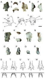
Figure 1. Alhuenia eduardotonnii (MACN SC 1439, holotype), distal end of right humerus in A,F, anterior; B,G, lateral; C,H, medial; D,I, posterior; and E,J, distal views. K-Q, anterior view of right humerus of selected gruiforms and cariamiforms, K, Parvigrus sp. (redrawn from Mayr, 2013a); L, Alhuenia eduardotonnii nov. sp.;
M, Aramus guarauna; N, Balearica sp.; O, Psophia crepitans; P, Rallus sanguinolentus; Q, Cariama cristata. Abbreviations. aa, anterior articular ligament; bf, brachial fossa; dc, dorsal condyle; ent, entepicondylar process; fp, flexor process; htg, humerotricipital groove; mcr, scar for the m. carpi radialis; of, olecranal fossa; vc, ventral condyle. 1, attachment of the tendon of the m. carpi radialis represented by low and elongate scar; 2, prominent attachment for anterior articular ligament; 3, centrally located brachial fossa; 4, transversely expanded distal end of humerus; 5, ventrally extended flexor process; 6, ventrally slanted distal end of humerus. Scale bar: A-J, 1 cm.; K-Q, not to scale.
1694433120323.png
 
Falconiformes Sharpe, 1874
Falconidae Leach, 1820
Genus Caroohierax nov. gen.

Diagnosis. Very small falconid diagnosed on the basis of the following unique combination of characters: 1- subtriangular cross-section of metatarsal shaft; 2- well-delimited and deep extensor groove forming a canal; 3- poorly defined surface of the impression for the m. abductor digiti II; 4- posterior surface of the shaft lacking prominent plantar crests; 5- metatarsal II trochlea strongly distally extended and transversely narrow, wellseparated from the posteromedial wing; 6- posteromedial wing strongly medially oriented and proximally extended; and 7- metatarsal trochlea IV laterally delimited by a wide, flattened and obliquely oriented lateral surface.

Etymology. Caroo, from the Aonikenk language, meaning carancho (in spanish) or caracara; hierax, from the Greek, meaning falcon.

Type and only included species. Caroohierax rapoporti nov. sp.

Caroohierax rapoporti nov. sp.

Diagnosis. The same as for genus by monotypy.

Holotype. MACN SC 1400, distal end of left tarsometatarsus with somewhat eroded distal trochleae

Locality and horizon. Loma de la Lluvia fossil site, Pinturas river, Santa Cruz province, Argentina. The specimen comes from beds belonging to the Early-Middle Miocene Pinturas Formation.

Etymology. In honour of Eduardo Rapoport (1927-2017) one of the most prominent Argentine biologists who greatly contributed to the knowledge of the biogeography and ecology of the southern cone.

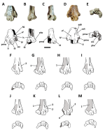
Remarks. In spite of the poor preservation of the only known Caroohierax specimen, it strongly departs from the morphology of other Miocene falconids. In fact, it shows a unique combination of features that make it difficult to assign it to any falconid subclade (see Becker, 1987; Cenizo et al., 2013; Noriega et al., 2011). It shows several features similar to that of Herpetotheriinae, including a subtriangular cross-section of the shaft, and a prominent and posteriorly oriented wing on metatarsal trochlea IV. Furthermore, Caroohierax differs from Falconini and Polyborini in that the extensor groove of digit IV lacks any sign of a double opening (Cenizo et al., 2013), the opening of the distal interosseus canal is located within the lateral intertrochlear incisure and lacks a proximally extended posterior notch. Additionally, Caroohierax differs from polyborines in the different shape and proportions of the distal trochleae, as well as in the absence of rugosities and pits delimiting the distal end of the surface for the m. abductor digiti II. However, in spite of the similarities noted above, Caroohierax differs from herpetotheriines in having a small and poorly defined surface for the m. abductor digiti II (such a wide surface is also widespread in falconines), in lacking well-defined plantar ridges and strongly excavated anterior and posterior metatarsal surfaces, and in lacking a wide and deep impression for the m. adductor digiti II . The shape of trochlea II in Caroohierax differs from that of herpetotheriines such as Thegornis, Herpetotheres and Micrastur in having a narrow and distally extended articular body (see Noriega et al., 2011).
Caroohierax differs from the basal falconid Antarctoboenus in having a deep and well-defined impression for the m. adductor digiti II , in the distal extension of trochlea II, in having narrow intertrochlear spaces, a narrow and deep extensor groove, and in stouter distal trochleae, among other features (Cenizo et al. , 2013).

Fred


Figure 1. Caroohierax rapoporti (MACN SC 1400, holotype) distal end of left tarsometatarsus in A, anterior; B, lateral; C, medial; D, posterior; and E, distal views. F-M, left tarsometatarsus of selected falconids in anterior view, F, Antarctoboenus carlini; G, Herpetotheres cachinans (Herpetotheriinae); H, Micrastur semitorquatus
(Micrasturinae); I, Polyborus plancus (Polyborini); J, Spiziapteryx circumcincta (Polyborini); K, Falco femoralis (Falconini); L, Caroohierax rapoporti; M, Thegornis musculosus (Herpetotheriinae). Abbreviations. adII, impression of the m. adductor digiti II; eg, extensor groove of digit IV; ls, lateral surface delimiting the trochlea IV; mfI, metatarsal I fossa; oic, distal opening of interosseous canal; plw, posterolateral wing; pmw, posteromedial wing of trochlea II; vf, distal vascular foramen. 1, expanded, deep and well-defined surface for the m. abductor digiti II; 2, wide lateral surface proximally delimiting trochlea IV; 3, double opening of distal vascular foramen; 4, subrectangular-shaped and transversely narrow body of trochlea II. In F-M, the surface shaded light grey represents the surface for the m. abductor digiti II. Scale bar: A-E, 2 mm.; F-M, not to scale. F-K, redrawn from Cenizo et al. (2013) and personal inspection of specimens; M, redrawn from Noriega et al. (2011).
1694434337588.png
 
Herpetotheriinae Lesson, 1842
Genus Thegornis Ameghino, 1895

Thegornis spivacowi nov. sp.

Diagnosis. Very small Thegornis species distinguishable by the following combination of characters: 1- tarsometatarsus in distal view with metatarsal IV trochlea having the posterior wing well-separated from the main body of the trochlea, resulting in a sigmoidal outer margin of the trochlea IV; 2- trochlea II robust and rounded, with a distal notch separating the anterior articular surface from the posterior wing; and 3- proximal ridge resulting in a medial prominence dorsal to the articular surface of trochlea II.

Holotype. MACN-SC 1407, distal end of right tarsometatarsus

Paratypes. MACN-SC 1390, distal end of left tarsometatarsus lacking II trochlea; MACN-SC 1393, distal end of left tarsometatarsus lacking IV trochlea (Figure 15 J-M); MACN-SC 1396, distal end of left tarsometatarsus lacking IV trochlea (Figure 15 Q-S); MACN-SC 4510, distal end of right tarsometatarsus lacking IV trochlea (Figure 15 G-I);
MACN-SC 4511, distal end of left tarsometatarsus with partially eroded distal trochleae.

Locality and horizon. Loma de la Lluvia fossil site, Pinturas river, Santa Cruz province, Argentina. The specimen comes from beds belonging to the Early-Middle Miocene Pinturas Formation. The specimen MACN-SC 4511 comes from the Portezuelo Sumich
locality (see Locality and horizon. Loma de la Lluvia fossil site, Pinturas river, Santa Cruz province, Argentina. The specimen comes from beds belonging to the Early-
Middle Miocene Pinturas Formation. The specimen MACN-SC 4511 comes from the Portezuelo Sumich locality (see Bown and Larriestra, 1990).

Etymology. Honouring José Boris Spivacow (1915-1994), editor of EUDEBA and Centro Editor de América Latina. Due to his efforts, more than 5000 titles were published, and without any doubt his projects have been of capital importance for Argentine cultural heritage.

Remarks. The material belonging to T. spivacowi nov. sp. was previously identified as a caracarine falconid by Chiappe (1991). On the contrary, Thegornis spivacowi nov. can be nested among herpetotheriine falconids based on having the following combination of features, including the roughly “H” shaped metatarsal shaft with a prominent lateral anterior ridge, resulting in a nearly subtriangular general contour and in a very wide and extended impression of the m abductor digiti II , metatarsal II trochlea with posteromedial wing slightly medially oriented, rounded and prominent articular surface of trochlea II, the lateral trochlear rim of metatarsal III trochlea more posteriorly extended than the medial one, relatively thin trochlea IV and extensive posterior wing of metatarsal IV trochlea (Noriega et al., 2011; Cenizo et al., 2013). Thegornis spivacowi comfortably fits within the genus Thegornis, with which it shares a tarsometatarsus with the anterior surface excavated and delimited by sharp ridges intermediate in size between Micrastur and Herpetotheres, an anterolateral margin of shaft elevated as a prominent border, resulting in a roughly subtriangular contour of the shaft, a relatively low posterior lateral metatarsal ridge, large and deep metatarsal I fossa that it is proximally delimited by a prominent ridge, trochlea II slightly grooved, and posteromedial wing of trochlea II relatively thin, well-defined and
strongly medially tilted. Ameghino (1895) coined the falconid genus Thegornis with the aim of including the Santacrucian species Thegornis musculosus and T. debilis, which were distinguished mostly by their disparate size. Later, Thegornis was assigned to Accipitridae (Brodkorb, 1964; Agnolín, 2006b). However more recently, Noriega et al. (2011) based on newly collected specimens, including a nearly complete skeleton, referred Thegornis to the family Falconidae, Subfamily Herpetotherinae. They also sustained that the size difference between T. debilis and T. musculosus may fit the sexual dimorphism present in extant falconids and considered that the only valid species is T. musculosus, a criterion with which I concur. T. spivacowi comes from younger beds than those which yielded T. musculosus. In spite of their similarities, T. spivacowi is a smaller species that can be distinguished by details of the distal trochleae of tarsometatarsus. These include a different shape of metatarsal IV trochlea with a well-defined posterior wing and strongly sigmoidal lateral margin. The prominent and rounded metatarsal II articular surface of T. spivacowi nov. resembles the condition of buteonine accipitrids (Noriega et al., 2011). However, in contrast with Thegornis musculosus the ventral margin of the trochlea II is not complete, with the anterior articular surface being separated from the posteromedial wing by a distal notch.

Remark by me: Thiks solves the "mystery" raised by me in the thread Thegornis sosae sp. nov.

Fred


Figure 1. Thegornis spivacowi. A-F, distal end of left tarsometatarsus lacking II trochlea (MACN-SC 1390, paratype) in A, anterior; B, lateral; C, medial; D, posterior; and E, distal views; F, cross-section of the shaft. G-I, distal end of right tarsometatarsus lacking IV trochlea (MACN-SC 4510, paratype) in G, anterior, H, posterior, and I, distal views; J-M, distal end of left tarsometatarsus lacking IV trochlea (MACN-SC 1393, paratype) in J, anterior; K, medial; L, posterior; and M, distal views; N-P, distal end of left tarsometatarsus with partially eroded distal trochleae (MACN-SC 4511, paratype) in N, anterior; O, posterior; and P, distal views; Q-S, distal end of left tarsometatarsus lacking IV trochlea (MACN-SC 1396, paratype) in Q, anterior; R, posterior; and S, distal views. Scale bar 5 mm.
1694435589025.png
 
Strigiformes Wagler, 1830
?Sophiornithidae Mourer-Chauviré, 1987
Genus Enskenia nov. gen.

Diagnosis. Medium-sized owl diagnosed on the basis of the following unique combination of features: 1- distal trochleae forming an opened “U” in distal view; 2- metatarsal trochlea IV small, poorly excavated, and laterally deflected; 3- metatarsal
trochlea III proximodistally short (especially in posterior view), with a well-defined and proximally raised margin; 4- outer rim of metatarsal III trochlea obliquely oriented; 5- distal margin of metatarsal III rochlea distally concave; 6- medial metatarsal plantar ridge prominent and acute; and 7- distal vascular foramen wide and distally ositioned, distally surrounded by a concave pit.

Etymology. Ensken, meaning “night” in Aonikenk language.

Type and only known species. Enskenia galeanoi nov. sp.

Enskenia galeanoi nov. sp.

Diagnosis. The same as for genus by monotypy.

Holotype. MACN SC 1609, distal end of right tarsometatarsus lacking trochlea II.

Paratypes. MACN SC 1421, distal end of right tarsometatarsus, lacking II and IV trochleae MACN SC 1430, trochlea III of right tarsometatarsus.

Locality and horizon. Loma de la Lluvia fossil site, Pinturas river, Santa Cruz province, Argentina. The specimen comes from beds belonging to the Early-Middle Miocene Pinturas Formation.

Etymology. Honouring Eduardo Galeano (1940-2015), born in Uruguay, one of the most preeminent Latin American writers.

Remarks. In spite of being poorly preserved, the tarsometatarsus of Enskenia nov. gen. shows a unique combination of characters that allows for recognizing its affinities among strigiforms. In contrast to crown-Strigiformes, Paleoglaucidae, Protostrigidae, and Ogygoptyngidae it shows poorly excavated anterior and posterior metatarsal grooves (despite being incompletely preserved, the anterior surface of the bone in most owls shows a concave surface that reaches the level of the distal vascular foramen), and a poorly arched distal end of the bone (Vickers Rich and Bohaska, 1981; Mourer-Chauviré, 1987, 1994; Mayr, 2016b), a combination of characteristics that distinguish the extinct clade Sophiornithidae. Furthermore, Enskenia differs from Protostrigidae, Paleoglaucidae, and Ogygoptyngidae in having a robust and transversely wide tarsometatarsus shaft (Martin and Black, 1972; Mayr, 2016b; Mourer-Chauviré, 1987, 1994; Peters, 1992; Vickers Rich and Bohaska, 1981). In spite of little overlapping material, Enskenia differs from the enigmatic Primoptynx in having a deeply grooved trochlea III with prominent condyles, and trochlea IV not strongly transversely compressed (Mayr et al., 2021). Moreover, the tarsometatarsus of Enskenia appears to be much more robust than the elongate one of Primoptynx (Mayr et al., 2020a).
Enskenia is very similar in shape and size to that of other sophiornithids, including the genera Sophiornis and Berruornis (Mourer-Chauviré, 1987, 1994). Comparisons with the genus Sophiornis are difficult because of few overlapping characteristics. However, Enskenia differs in having the distal end of the bone transversely narrower, and in having a more proximally positioned distal vascular foramen, which is far from the proximal margin of the trochlea III (a condition shared with Berruornis ; Mourer-Chauviré, 1994). This contrasts with Sophiornis, where the foramen is located on level with the trochlea III and is very close to its proximal margin. Furthermore, in Enskenia the metatarsal I fossa is shallower and narrower than in Sophiornis (Mourer-Chauviré, 1987).
Enskenia differs from Berruornis in having metatarsal trochlea III strongly raised from the metatarsal shaft and with deep trochlear rims (Mourer-Chauviré, 1994). The trochlea IV in Enskenia is strongly laterally oriented, a condition that departs from that of Berruornis .

Fred


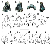
Figure 1. Enskenia galeanoi. A-I, distal end of right tarsometatarsus lacking trochlea II (MACN SC 1609, holotype) in A,G, anterior; B, proximal; C,H, distal; D,I, posterior; E, medial; and F, lateral views. J-N, distal end of right tarsometatarsus, lacking II and IV trochleae (MACN SC 1421, paratype) in J, anterior; K, proximal; L., distal; M, posterior; and N, medial views. O-R, trochlea III of right tarsometatarsus (MACN SC 1430, paratype) in O, anterior; P, posterior; Q, distal; and R, medial views. S-W, distal end of right tarsometatarsus of selected strigiforms in anterior (top) and distal (bottom) views: S, Berruornis orbistanqui; T, Sophiornis quercynus; U, Enskenia galeanoi; V, Eostrix gulottai; W, Asio flammeus. The lines at the bottom figures represent the main axis of the body of distal trochlea in distal view when compared with the horizontal plane. This evidences the much wider angle present in sophiornithids (S-U) when compared with other strigiforms. Abbreviations. dvf, distal vascular foramen; iof, interosseous opening; lpr, lateral plantar ridge; mpr, medial plantar ridge; mtI, metatarsal I fossa; plw, posterolateral wing of metatarsal IV trochlea. S-T, redrawn from Mourer-Chauviré (1994); V, redrawn from Mayr (2016). Scale bar: 5 mm.
1694441314030.png
 
Coraciiformes sensu Mayr, 1998
Coracii Wetmore and Miller, 1926
Coraciidae Batsch, 1788
Genus Chehuenia nov. gen.

Diagnosis. Coraciid diagnosed on the basis of the following unique combination of characteristics: 1- very large trochlea II with a proximally extended medial wing; 2- posteromedial wing of metatarsal trochlea II strongly posteriorly extended; 3- poorly excavated posterior surface of trochlea II; 4- IV metatarsal trochlea only slightly shorter than trochlea III and the latter longer than II metatarsal trochlea; 5- transversely narrow IV metatarsal trochlea; 6- proximally positioned distal vascular foramen; and 7- deep longitudinal groove separating metatarsals II and III in posterior view.

Etymology. Cheuen, from the Aonikenk language: little bird.

Type and only included species. Chehuenia facongrandei nov. sp.

Chehuenia facongrandei nov. sp.

Diagnosis. The same as for genus by monotypy.

Holotype. MACN SC 4512, distal end of left tarsometatarsus.

Locality and horizon. The specimen comes from the Santa Cruz Formation (Early-Middle Miocene), at the well-known fossiliferous locality of Monte León (the label indicates “Estaca 3”), Monte León National Park, Santa Cruz province, Argentina

Etymology. facongrandei, honouring José Font “Facón Grande” (1883-1921), an Argentine worker and anarcho-syndicalist, killed during rural strikes in Patagonia by 1921.

Remarks. The tarsometatarsus of Chehuenia nov. gen. is referable to Coraciidae on the basis of the following unique combination of characteristics (Bourdon et al., 2016; Degrange et al., 2021; Mayr, 2022; Mayr and Mourer-Chauviré, 2000; Mourer-
Chauviré, 1999; Mourer-Chauviré et al., 2013): short, wide and flattened distal end of the tarsometatarsus, the anterior surface lacks a wellmarked extensor groove, trochleae II and IV shorter than trochlea III, trochlea II reaches farther distally
than trochlea IV (trochleae III and IV are subequally extended distally in Geranopteridae; Mayr and Mourer-Chauviré, 2000; Mourer-Chauviré, 1999), deep distal interosseous groove between trochleae III and IV, and enlarged distal vascular foramen.
In spite of being represented by several fossils in the past, the fossil record of Coraciidae is now restricted to the extinct genus Miocoracias and the still living genera Coracias and Eurystomus (Mourer-Chauviré et al., 2013). Most other taxa previously referred as being fossil coraciids resulted as being part of other coraciiform lineages (Mayr, 2009; Mayr and Mourer-Chauviré, 2000; Mourer-Chauviré, 1999;
In spite of being represented by several fossils in the past, the fossil record of Coraciidae is now restricted to the extinct genus Miocoracias and the still living genera Coracias and Eurystomus (Mourer-Chauviré et al., 2013). Most other taxa previously referred as being fossil coraciids resulted as being part of other coraciiform lineages (Mayr, 2009; Mayr and Mourer-Chauviré, 2000; Mourer-Chauviré, 1999; Mourer-Chauviré et al. , 2013). Comparisons between Chehuenia, Miocoracias, Coracias, and Eurystomus indicate that the former is a distinctive taxon. In Chehuenia the distal vascular foramen is proximally positioned and is proximodistally elongate, being similar in both aspects to the extinct genus Miocoracias, and different from the condition of living genera Coracias and Eurystomus (Mourer-Chauviré et al., 2013). Its metatarsal trochlea IV is only slightly distally extended than trochlea III, in contrast to the shorter condition shown by Miocoracias and Coracias. IV metatarsal trochlea in
Chehuenia is transversely narrow and subrectangular in contour, contrasting with the subquadrangular condition present in Miocoracias, Coracias, and Eurystomus. Its metatarsal IV shows a welldeveloped posterior wing that is more developed than in Miocoracias and Eurystomus, whereas in Coracias this wing is nearly absent. Metatarsal III trochlea of Chehuenia shows well defined and prominent rims separated by a deep groove as in Miocoracias, and contrasting with Coracias and
Eurystomus. II metatarsal trochlea is thicker than in Miocoracias and Coracias, and its posteromedial wing is very well-developed, and is as posteriorly extended as in Coracias. The posterior opening of the distal vascular foramen in Chehuenia is ovoidal in contour, as in Coracias and Eurystomus, but contrasting with the more elongate condition present in Miocoracias. In distal view the trochleae form a “U” shaped opened arch, more marked than in Eurystomus and Coracias.
As remarked by the comparisons made above, the position of Chehuenia within Coraciidae is not clear. As indicated in the descriptive section, there are some differences with extant members of Coraciidae. In contrast to extant members of the clade, the trochlea metatarsi III is distally splayed (Mourer-Chauviré et al., 2013). On the other side, some similarities are noted with Miocoracias from the Early Miocene of France, namely, the well developed posterior wing of metatarsal IV trochlea and arched trochleae in distal view (see Mourer-Chauviré et al., 2013). The incomplete nature of the available specimen does not allow for refining the phylogenetic position of Chehuenia among coraciids. The only previous fossil record of fossil coraciiforms in South America is Ueekenkcoracias tambussiae, from the Eocene of Chubut province,
Patagonia (Degrange et al., 2021; but see Mayr, 2022). Chehuenia differs from Ueekenkcoracias in having metatarsal trochlea IV more distally extended than trochlea II, and in that the II trochlea is not strongly medially deflected.

Fred


Figure 1. Chehuenia facongrandei (MACN SC 016, holotype) distal end of left tarsometatarsus in A,E, anterior; B,F, posterior; C,G, distal; and D,H, lateral views. I, Miocoracias chenevali, distal end of left tarsometatarsus in distal view. J-O, left tarsometatarsus in anterior view of selected coraciiforms: J, Chehuenia facongrandei; K, Coracias garrulus (Coraciidae); L, Miocoracias chenevali; M, Eurystomus glaucurus (Coraciidae); N, Geranopterus alatus (Geranopteridae); and O, Ueekenkcoracias tambussiae. Abbreviations. ig, interosseous groove; plw, posterolateral wing; vf, distal vascular foramen; II-IV, corresponding number of distal trochlea; 1, deep and well-defined anterior interosseous groove between metatarsals III and IV; 2, distal trochlea II more distally extended than trochlea IV, being close to the distal level of trochlea III; 3, proximally positioned trochlea IV; 4, distally splayed trochlea III. Scale bar: A-D, 5 mm; J-O, not to scale. I, L, redrawn from Mourer-Chauviré et al. (2013); K,M, redrawn from Mourer-Chauviré (1999); N, redrawn from Mayr and Mourer-Chauviré (2000);
O, redrawn from Degrange et al. (2021).
1694447815194.png
 
Passeriformes Linnaeus, 1758
Tyranni Wetmore and Miller, 1926
Indeterminate genus and species

Referred material. MACN SC 3300, incomplete distal end of right tarsometatarsus MACN SC 3303, incomplete distal end of right tarsometatarsus; MACN SC 3304,
incomplete distal end of left tarsometatarsus; MACN SC 3305, incomplete distal end of right tarsometatarsus

Locality and horizon. Loma de la Lluvia fossil site, Pinturas River, Santa Cruz province, Argentina. The specimens come from beds belonging to the Early-Middle Miocene Pinturas Formation.

Remarks. The specimens described here are referred to passeriforms because of the combined presence of distal trochlea in the same plane when viewed distally, with III trochlea notably larger than IV and IV, wide intertrochlear grooves, and the lateral plantar ridge not prominent (Hamon, 1964; Mayr and Manegold, 2004; see also Manegold, 2008). Furthermore, in spite of their incomplete nature, the specimens may be included among Tyranni suboscines by having the scar for metatarsal I not well-raised from the shaft, trochlea II relatively small, trochlea III slightly distally expanded, and trochlea IV somewhat laterally inflected and excavated (the later shared with Menurae; Ballmann, 1969; Boles, 1995a,b; Hamon, 1964; Manegold, 2008). Moreover, as in most Tyranni, the distal end of the bone proximal to the trochleae is strongly flattened (Pycraft, 1906).
Among Tyranni, the phylogenetic position of available specimens is not certain. They differ from Dendrocolaptidae because trochlea IV is much shorter and smaller than trochlea III and because the distal end of the bone does not suddenly broadens transversely (Feduccia, 1973; Manegold, 2008; Maurício et al. , 2012). In contrast to Thamnophilidae and “Furnariidae” trochlea IV is distinctly grooved (shared with Rhinocryptidae and Formicariidae; Feduccia and Olson, 1982; Maurício et al., 2012).
Trochlea III shows a well-defined and deep groove, as occurs in Dendrocolaptidae, Rhinocryptidae and Formicariidae, contrasting with “Furnariidae” and Tyrannidae (see Feduccia and Olson, 1982; Manegold, 2008; Maurício et al. , 2012). In contrast to Dendrocolaptidae and “Furnariidae” trochlea II does not exceed the anterior surface of the tarsometatarsus (Manegold, 2008). In most of the above mentioned features, available specimens are similar to Formicariidae, but a taxonomic referral is far from certain. It is preferred here to leave the specimens as indeterminate Tyranni.

Fred


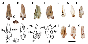
Figure 1. Tyranni indet. A-C, Morphotype 3 (MACN SC 3304), incomplete distal end of left tarsometatarsus in A, anterior; B, posterior; and C, distal views; D-E, Morphotype 1 (MACN SC 3303), incomplete distal end of right tarsometatarsus in D, anterior; and E, posterior views; F-H, Morphotype 2 (MACN SC 3300), incomplete distal end of right tarsometatarsus in F, lateral; G, anterior; and H, posterior views; I-K, Morphotype 3 (MACN SC 3305), incomplete distal end of right tarsometatarsus in I, anterior; J, posterior; and K, medial views. Abbreviations: eg, outer extensor groove; ep, extensor pit; fp?, possible flexor pit; ig, intermetatarsal grooves; it, intertrochlear groove; mtI, fossa for metatarsal I; vf, distal vascular foramen; IV, fourth metatarsal trochlea. Scale bar: 2 mm.
1694448444323.png
 

Users who are viewing this thread

Back
Top