Systematic palaeontology
Class Aves Linnaeus, 1758
Order Anseriformes Wagler, 1831
Family Anatidae Leach, 1819
Genus
Manuherikia Worthy, Tennyson, Jones, McNamara and Douglas, 2007
Remarks: The new species is referred to the genus
Manuherikia because it has the unique combination of generic features described by Worthy et al. (2007), namely, for the humerus:
- fossa pneumotricipitalis dorsalis broad, markedly undercuts the caput over its width from adjacent to the tuberculum dorsale to incisura capitis;
- the insertion of m. scapulohumeralis cranialis (wrongly equated with crus dorsale fossae by Worthy et al., 2007) is large and prominent, extending distally to a point level with where the crista bicipitalis joins the shaft;
- the tuberculum dorsale elevated, ovate and aligned with shaft;
- the fossa m. brachialis is deep; the facet for the attachment for the anterior articular ligament is well-defined, as long as wide, elevated and tilted distally.
To these we add the following humeral features that characterise all members of the genus:
- humerus with fossa pneumotricipitalis ventralis forming a closed (not highly pneumatic), deep tapered pocket extending craniodorsal to the crus dorsale fossae to undercut the caput;
- the tuberculum ventrale projects caudally over the fossa pneumotricipitalis ventralis but does not overhang it distally;
- a well-defined capital shaft ridge proximally extending to a point just ventral to tuberculum dorsale;
- the crista deltopectoralis is slightly concave dorsally, angulus cristae at its mid-length point, and about 30% of its length extends distal to the crista bicipitalis;
- the scar for the humeral anchor of m. scapulotriceps (= the scar for the m. latissimus dorsi posterioris, sensu Howard, 1929 and Woolfenden, 1961) is distal to and in line with the tuberculum dorsale (pectoral attachment), not displaced to be entirely dorsal to the
tuberculum, as defines ‘Oxyurini’ sensu Woolfenden (1961, p. 20);
- the insertion scar of m. latissimus dorsi cranialis proximally is caudally-separated from the crista deltopectoralis, and as it extends distally converges with and passes closely caudal to the end of the crista and then extends as a linear prominence on the dorsal shaft;
- the shaft is slightly sigmoid, narrowing distally with least shaft width near distal end;
- distally, the epicondylaris dorsalis is slightly prominent dorsally adjacent to the condylus dorsalis, but a distinct processus supracondylaris dorsalis is lacking;
- the origin of m. extensor metacarpi radialis is proximal to the epicondylaris dorsalis and extends from the dorsal facies onto the cranial facies as a slightly raised rugose area;
- the fossa m. brachialis is ovate, wider proximally where extends over half of shaft width,narrows distally to be deepest just proximal to tuberculum supracondylare ventrale and is narrowly separated from the ventral margin;
- the processus flexorius is directed caudally such that the epicondylus ventralis is more prominent ventrally;
- in cranial/caudal aspect, the processus flexorius has about equal distal extent as the condylus dorsalis and condylus ventralis;
- the pit for the origin of m. extensor metacarpi ulnaris (flexor carpi ulnaris; that furthest from the facet for the anterior ligament), is reduced (much shallower than the adjacent pit for the origin of m. pronator profundus), a characteristic of ‘Oxyurini’ (Woolfenden,
1961: 20).
Manuherikia primadividua nov. sp.
LSID of new species: urn:lsid:zoobank.org:act:3944149D-E74B-4931-83F6-20ACAEA53EE1.
Derivation of the name: from prima, first and dividua, divisible or divided, feminine nominative, to acknowledge this small duck provides the first evidence of a division in the St Bathans Fauna.
Holotype: CM 2013.18.1181, right humerus
Paratypes: CM 2013.18.692 p+d parts L humerus; CM 2013.18.706 L hum; CM 2013.18.710 p+d parts L hum; CM 2013.18.1179 R hum (little crushed proximally); CM 2013.18.1187 d+s R hum; CM 2013.18.1188 dR hum.
Type Locality: Mata Creek, Site 9, near St Bathans, Otago, New Zealand. The outcrop is on the western bank of Mata Creek. Coordinates: 44° 52.8288’ S, 169° 50.4162’ E. NZ Fossil Record File Number H41/f0122.
Referred specimens (* exemplary ones):
Manuherikia River, Bed HH6: CM 2013.18.420, frags R ulna; CM 2013.18.421, pL ulna; CM 2013.18.423, L hum; CM 2013.18.440, L hum; CM 2013.18.759, L ulna (2 pts); CM 2013.18.760, dL ulna; CM 2013.18.761, R cor; CM 2013.18.762, R hum; CM 2013.18.763, L hum; CM 2013.18.766, R cor; CM 2013.18.1331, L fem; CM 2013.18.1332, dR tib.
Manuherikia River, Bed HH7: CM 2013.18.5, cranium; CM 2013.18.6, dL fem*; CM 2013.18.7, dR hum; CM 2013.18.8, R tmt; CM 2013.18.9, dL tib; CM 2013.18.11, dL tib*; CM 2013.18.12, L&R rami mandibles; CM 2013.18.13, dL tib*; CM 2013.18.14&15, pR + dR fem with medullary bone; CM 2013.18.16, L tmt; CM 2013.18.17, d+sR tmt; CM 2013.18.18, sL hum; CM 2013.18.19, dR tib; CM 2013.18.20, p+sL hum; CM 2013.18.21, dR tib; CM 2013.18.22, pR rad; CM 2013.18.24, cranial pt R cor; CM 2013.18.25, L ulna.
Manuherikia River, Bed HH8: CM 2013.18.1578, pR cmc*.
Manuherikia River, 100 m downstream of bridge: NMNZ S.53301, pL hum.
Mata Creek, Site 4: CM 2017.37.644, R cor; CM 2017.37.645, sternal end L cor; NMNZ S.53256, R quad.
Mata Creek, Site 5: CM 2013.18.829, d+s R hum; CM 2013.18.830, dL hum; CM 2013.18.831, R ulna; CM 2013.18.832, R scap; CM 2013.18.833, pR tib (frags); CM 2017.37.358, cranial pt L cor; CM 2017.37.359, pR ulna; NMNZ S.53480, pL tmt.
Mata Creek, Site 9, main bonebed (unless otherwise specified): CM 2013.18.623, R MII.1; CM 2013.18.625, L scap; CM 2013.18.626, pR tmt; CM 2013.18.627, pL fem*; CM 2013.18.628, pR tib*; CM 2013.18.629, R fem (-ball)
; CM 2013.18.631, L scap; CM 2013.18.654, R scap; CM 2013.18.669, R scap; CM 2013.18.677, dR tib; CM 2013.18.681, R cor*; CM 2013.18.685, R dentary; CM 2013.18.687, pL tib*; CM 2013.18.688, R cor*; CM 2013.18.1183, dL tib*; CM 2013.18.1184, dR fem*; CM 2013.18.1185, dL fem; CM 2013.18.1186, dR fem; CM 2013.18.1189, dR tmt*; CM 2013.18.1190, L tmt*; CM 2013.18.1191, R tmt*; CM 2013.18.1192, R cor*; CM 2013.18.1193, R cor; CM 2013.18.1194, L cor*; CM 2013.18.1198, R ulna*; CM 2013.18.1199, L ulna*; CM 2013.18.1200, R scap; CM 2013.18.1203, R rad; CM 2013.18.1204, L rad; CM 2013.18.1205, L rad; CM 2013.18.1239, pL tmt*; CM 2013.18.1243, R rad; CM 2013.18.1245, L quad; CM 2013.18.1247, L tmt*; CM 2013.18.1251, R scap; CM 2013.18.1253, pL rad; CM 2013.18.1255, dR ulna*; CM 2013.18.1284, R scap; CM 2013.18.1285, dR ulna; CM 2013.18.1286, L scap; CM 2013.18.1287, L scap; CM 2013.18.1288, L MII.1; CM 2013.18.1293, dR hum; CM 2013.18.1299, R scap; CM 2013.18.1301, L ramus mand; CM 2013.18.1330, anterior sternum; CM 2013.18.1351 (Lower Shell Layer, 102 cm below main bone layer), pR hum; CM 2013.18.1361 (10-20 cm above Upper Shell Layer), dR tib*; NMNZ S.53552, cranium; NMNZ S.53558, L cmc*; NMNZ S.53559, pelvis fragment; NMNZ S.53562, L ulna (-d); NMNZ S.53564, L hum; NMNZ S.53565, L cor; NMNZ S.53566, dL rad; NMNZ S.53567, L cor.
Mata Creek, Site 10A: CM 2017.37.122, pR rad; CM 2017.37.124, synsacrum; CM 2017.37.125, pL fem; CM 2017.37.127, R cor; CM 2017.37.128, R ulna; CM 2017.37.693, L fem*; NMNZ S.53895, cran pt L cor (in block).
Mata Creek, Site 10E: CM 2017.37.654, L quad.
Stratigraphy and age: Bannockburn Fm., Manuherikia Group, lower Miocene (19–16 Ma). A maximum of 2.6 m of green to olive-green clay is exposed below alluvial gravels at the south or downstream end of the bank in which the beds dip north at ~15 degrees. Three fossil bone bearing horizons were identified in the section measured at the south end of the outcrop: the ‘Lower Shell Layer’, 300–320 mm above the base, with densely packed mollusc fragments, fish bones, and uncommon bird fragments; the ‘Upper Shell Layer’, 820–860 mm above the base, marked by mollusc fragments up to 40 mm long, and fewer bones; and the ‘Main bone layer’, the source of the holotype, an olive green clay that is slightly more silty than bounding layers, marked by mm-sized fragments of calcified tubes showing as white spots in trowelled section, and rare molluscs and bird bones, 1380–1800 mm above the base (Table 1).
Measurements (in mm): Holotype L humerus: length 65.8 mm, proximal width 13.7 mm, minimum SW 3.9 mm, maximum DW 8.4 mm. Other specimens: see Table 3.
Diagnosis: A species of
Manuherikia larger than
M. minuta and smaller than
M. lacustrina, with humeri differentiated from them as follows (their state in brackets) by: capital shaft ridge is rounded and broad in section adjacent to the tuberculum dorsale (compressed narrow); origin of the dorsal head of m. humerotriceps is not deeply impressed on the area dorsal and distal to the crus dorsale fossae (deeply impressed forming a marked sulcus so that the scar for insertion of the m. scapulohumeralis cranialis straddles an acute crest extending distal to the crus dorsale fossae); carpometacarpi differ by a shallower fossa infratrochlearis; stouter proportions; relatively longer, more cranially directed processus extensorius.
Fred
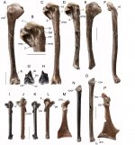
Figure 1. Pectoral elements of small species of
Manuherikia.
Manuherikia primadividua nov. sp.: Holotype R humerus CM 2013.18.1181 in cranial (A), caudal (B,C), and dorsal (F) aspect; L carpometacarpus NMNZ S.53558 in dorsal (J) and ventral (L) aspect; R ulna CM 2013.18.1198 in cranial aspect (O); R coracoid CM 2013.18.688 in dorsal aspect (P); compared to
M. minuta R humerus NMNZ S.53563 in caudal (D) and cranial (E) aspect; dR humerus CM 2017.37.195 in cranial (G) and caudal (H) aspect; L carpometacarpus NMNZ S.53456 in dorsal (I) and ventral (K) aspect; L coracoid CM 2013.18.1232 in dorsal
view (M); R ulna NMNZ S.53467 in cranial view (N). Abbreviations: 1, diagnosis character 1, capital shaft ridge; 2, diagnosis character 2, sulcus for origin of the dorsal head of m. humerotriceps; 3, diagnosis character 3, fossa infratrochlearis; 4, diagnosis character 4, processus extensorius; am, angulus medialis; ams, attachment of the humeral anchor of m. scapulotriceps; c, caput; cd, condylus dorsalis; cod, cotyla dorsalis; cov, cotyla ventralis; crd, crista deltopectoralis; csr, capital shaft ridge; cv, condylus ventralis; fac, facies articularis clavicularis; fb, fossa m. brachialis; fpd, fossa pneumotricipitalis dorsalis; msc, scar for insertion of m. scapulohumeralis cranialis; n, notch; omm, os metacarpale minus; pa, processus alularis; pe, processus extensorius; pra, processus acrocoracoideus; tc, tuberculum carpale; td, tuberculum dorsale; tsv, facet on tuberculum supracondylare ventrale. Scale bars:
10 mm.
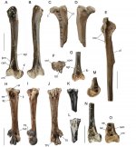
Figure 2. Leg elements of small species of
Manuherikia.
Manuherikia primadividua nov. sp.: R femur CM 2013.18.629 in caudal (A) and cranial (B) aspect; proximal L femur CM 2013.18.627 in cranial (C) and caudal (D) aspects; L tarsometatarsus, CM 2013.18.1247 in proximal (F), dorsal (H), medial (I), and plantar (J) aspects; distal R tibiotarsus, CM 2013.18.677 in medial (M), and cranial (O) aspect; distal R tibiotarsus,
CM 2013.18.1361 in cranial (N) aspect; compared to M. minuta distal L femur, CM 2017.37.24 in caudal aspect (G); proximal and shaft L tibiotarsus, CM 2013.18.400 in cranial aspect (E); and proximal L tarsometatarsus, NMNZ S.53077 in dorsal (K) and plantar (L) aspect. Abbreviations. c, caput; ccc, crista cnemialis cranialis; cd, condylus dorsalis; cf, crista fibularis; cm, condylus medialis; col, cotyla lateralis; com, cotyla medialis; csm, crista supracondylaris medialis; ct, crista trochanteris; ei, eminentia intercotylaris; fdl, canal for tendon of m. flexor digitorum longus; fi, fossa infracotylaris dorsalis; fp, fossa poplitea; fvd, foramen vasculare distale; ipm, insertion of puboischiofemoralis medialis; ire, medial impressio retinaculi extensoris; rel, lateral tuberositas retinaculi extensoris; rem, medial tuberositas retinaculi extensoris; se, sulcus extensorius; sgl, scar for the insertion of m. gastrocnemialis lateralis; tf, trochlea fibularis; TII, trochlea metatarsi II; TIV, trochlea metatarsi IV; ttc, tuberositas m. tibialis cranialis. Scale bars: 10 mm.