• Welcome to BirdForum, the internet's largest birding community with thousands of members from all over the world. The forums are dedicated to wild birds, birding, binoculars and equipment and all that goes with it.

    Please register for an account to take part in the discussions in the forum, post your pictures in the gallery and more.
ZEISS DTI thermal imaging cameras. For more discoveries at night, and during the day.

Marambiornopsis sobrali gen. et sp. nov. (1 Viewer)

Fred Ruhe

Well-known member
Netherlands
Piotr Jadwiszczak, Marcelo Reguero & Thomas Mörs, 2021

A new small-sized penguin from the late Eocene of Seymour Island with additional material of Mesetaornis polaris

GFF
. Online edition. doi:10.1080/11035897.2021.1900385

Free pdf: https://www.tandfonline.com/doi/pdf/10.1080/11035897.2021.1900385?needAccess=true


Abstract:

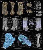
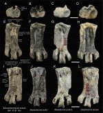
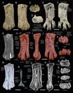
Here, we report on two tarsometatarsi assignable to relatively small-sized Eocene Antarctic penguins, housed in the palaeozoological collections of Naturhistoriska riksmuseet, Stockholm. The Priabonian fossils were collected by museum staff during two joined Argentinean and Swedish expeditions from the Submeseta Formation on Seymour Island, Antarctic Peninsula. One specimen represents a new early sphenisciform, Marambiornopsis sobrali gen. et sp. nov., the sixth small-sized tarsometatarsus-based penguin species known from the Antarctic Eocene. Micro-CT scanning revealed the presence of quite large and essentially empty metatarsal medullary cavities. The second fossil can unequivocally be assigned to Mesetaornis polaris. The specimen represents only the second record of this species and supposedly a relatively young bird. Micro-CT scanning showed that in M. polaris the metatarsal medullary cavities are less developed than in M. sobrali – the cortical and trabecular bone tissues left rather little room for significant hollow spaces. Both specimens also differ in overall density of their trabecular networks.

Enjoy,

Fred
 
Systematic palaeontology

CLASS AVES LINNAEUS, 1758
ORDER SPHENISCIFORMES SHARPE, 1891
FAMILY SPHENISCIDAE BONAPARTE, 1831

Remarks – Clarke et al. (2003: p. 3) postulated “that the name Spheniscidae’ be formally applied to the clade comprised of the most recent common ancestor of all extant penguins and all of its descendants”. Here, we use the name in its traditional context.

Genus Marambiornopsis gen. nov.

Type speciesMarambiornopsis sobrali sp. nov. from the Submeseta Formation, Seymour Island (Antarctic Peninsula).

Differential diagnosisMarambiornopsis differs from other Eocene penguins in possessing a unique combination of features listed below. Tarsometatarsus small (unlike in Anthropornis and Palaeeudyptes) and strongly elongated (unlike in Archaeospheniscus/Notodyptes). Both proximal vascular foramina small (like in Delphinornis and Marambiornis; both large in Archaeospheniscus/Notodyptes, medial one especially large in Mesetaornis). Plantar opening of medial proximal vascular foramen situated close to the medial margin of second metatarsal, just distal to medial hypotarsal crest, and separated from the main hypotarsal sulcus (distal or mesiodistal relative to the medial hypotarsal crest, and separated from the main hypotarsal sulcus in Delphinornis; located within the main hypotarsal sulcus, and separated from the medial margin by the hypotarsal crest in Marambiornis and Mesetaornis). Tuberosity for insertion of m. tibialis cranialis wide (clearly wider than in Marambiornis, Mesetaornis and Delphinornis). Hypotarsal sulcus for tendon of m. flexor hallucis longus distinct (unlike in Delphinornis), and only moderately wide (markedly narrower than in Marambiornis). Main hypotarsal sulcus (for tendon of m. flexor digitorum longus) located clearly abaxially relative to the main axis of the bone (like in Marambiornis and Mesetaornis, but said axis passes through the sulcus in Delphinornis). Medial hypotarsal crest clavate in proximal view (a variety of chisel-like shapes in Delphinornis, Marambiornis and Mesetaornis)/

Etymology – An existing name for a genus of Eocene fossil penguins (Marambiornis) combined with the Greek word ópsis for appearance, sight. Hence Marambiornopsis.

Marambiornopsis sobrali sp. nov.

Material – An isolated right tarsometatarsus; type specimen only.

Horizon and Locality – Level 38 within the Submeseta Formation (Submeseta II Allomember), locality NRM 2, Slope S Marambio Base, S 64° 14.778ʹ, W 056°37.169ʹ, Seymour Island, Antarctic Peninsula (Montes et al. 2013; Kriwet et al. 2016).

Age – Upper TELM 7, Priabonian, late Eocene (Douglas et al. 2014).

Holotype – NRM-PZ A542, Figs. 1 and 2A, E, I.

Etymology – Named in honour of Navy Sub-Lieutenant José Maria Sobral, Argentinean geologist and member of the Swedish South Polar Expedition 1901–03 that discovered the first Antarctic penguin fossils.

Diagnosis – As for genus.

Description – NRM-PZ A542 is a well-preserved tarsometatarsus. The specimen is 43.8 mm long (maximally), 17.4 mm wide proximally and 19.7 mm wide distally, whereas its midwidth amounts to 15.1 mm. Two modest proximal vascular foramina are more widely spaced plantarly than dorsally, and the lateral one appears to be larger. The canal connecting matching openings for each foramen is (as revealed by micro-CT scanning; Fig. 1O) arcuate. Distally to the plantar opening of the medial foramen there is a distinct elongate elevation of the bone margin. The medial intermetatarsal sulcus is shallow and very short, its lateral counterpart is much better developed and can be traced up to the intertrochlear incisure. The latter is also wide proximally. The distal vascular foramen is represented solely by its plantar opening. The wide tuberosity for the insertion of the m. tibialis cranialis appears to be tear dropshaped. The hypotarsus is not simplified, and three crests are distinguishable. The well-preserved medial crest is most prominent, and the other two are in varying degrees of damage. The curvature of the cross section of the main hypotarsal sulcus creates an almost perfect circular arc, with two crests partially overhanging a resulting passage. The surface for the origin of the m. abductor digiti IV is laterally framed by a low marginal crest. The second metatarsal is gently curved, its medial margin seems to be downright straight over a long distance of its length. The fourth metatarsal is straight. The third trochlea is clearly more robust than the other two. As revealed by micro-CT scanning (Fig. 1F–I, L–N, O–Q), the tarsal and proximalmost metatarsal as well as trochlear parts of the tarsometatarsus are rich in small, more or less interconnected, air pockets (hollow spaces within the 3D trabecular bony network; see Fig. 1Q), whereas the major parts of metatarsals are in this respect mostly dominated by large medullary cavities. The cavity inside the second metatarsal has the least variable diameter. The third metatarsal has the most spacious cavity, followed by the fourth and second metatarsals.

Fred


Figure 1. Type specimen (NRM-PZ A542) of Marambiornopsis sobrali gen. et sp. nov. (Aves: Sphenisciformes) from the Submeseta Formation, Seymour Island; A–E: the tarsometatarsus in dorsal, medial, plantar, proximal and distal views, F–I: its micro-CT coronal-plane slices, J–K: its annotated 3D surface model (micro-CT based), L–N: its inner-structure visualization focused on air pockets and medullary cavities (cortical bone transparent; micro-CT based), O–P: its inner-structure visualization in a cross-sectional view (cortical bone transparent; micro-CT based), Q: a longitudinal section (oblique lateral view) through the metatarsal III (cortical and trabecular bone opaque; micro-CT based); scale bar is 1 cm.

Figure 2. Late Eocene small-sized penguin tarsometatarsi from the Submeseta Formation, Seymour Island; features used in the diagnosis of Marambiornopsis marked; A, E, I: Marambiornopsis sobrali gen. et sp. nov., NRM-PZ A542 (holotype), B, F, J: Marambiornis exilis, IB/P/B-0490 (holotype), C, G, K: Mesetaornis polaris, IB/P/B-0278 (holotype, mirrored), D, H, L: Delphinornis larseni, IB/P/B-0062 (mirrored), all in proximal (A–D), dorsal (E–H) and plantar (I–L) views respectively;
scale bar is 1 cm.
 

Attachments

  • Marambiornopsis sobrali.jpg
    Marambiornopsis sobrali.jpg
    1.4 MB · Views: 1
  • Marambiornopsis sobrali-2.jpg
    Marambiornopsis sobrali-2.jpg
    1.2 MB · Views: 0
Last edited:
Genus Mesetaornis Myrcha et al., 2002

Diagnosis– According to Myrcha et al. (2002): “Strongly elongated tarsometatarsus with quite large foramen vasculare proximale mediale (smaller in Delphinornis and Marambiornis). Differs from Delphinornis in having the plantar opening of the foramen situated more medially (in relation to the main axis of the bone) than crista medialis hypotarsi. Tarsometatarsus differs from Marambiornis in having cristae intermediae hypotarsi separated by a relatively narrow groove”.

Species included– Monotypic, M. polaris from the Submeseta Formation, Seymour Island (Antarctic Peninsula).

Remarks– The genus is represented by two tarsometatarsi (IB/P/B-0278 and IB/P/B-0279b; Myrcha et al. 2002); however, only the former bone (Figs. 2C, G, K and Figs. 3D–F) has been originally assigned to a species (as a holotype). IB/P/B-0279b is very poorly preserved, but Myrcha et al. (2002) speculated that it could have represented another species. A few other specimens considered in this regard (Myrcha et al. 2002; Jadwiszczak 2006) are either isolated non-tarsometatarsal skeletal elements, devoid of “physical” ties to said tarsometatarsi, or are too incomplete for comparison. From this, it follows that the specimen described below is only the second specimen able to the sole named species of Mesetaornis.

Mesetaornis polaris Myrcha et al., 2002

New material– An isolated left tarsometatarsus NRM-PZ A824, partly in matrix.

Horizon and Locality– Level 38 within the Submeseta Formation (Submeseta II Allomember), locality NRM 2, Slope S Marambio Base, S 64° 14.778ʹ, W 056°37.169ʹ, Seymour Island, Antarctic Peninsula (Montes et al. 2013; Kriwet et al. 2016).

Age– Upper TELM 7, Priabonian, late Eocene (Douglas et al. 2014).

Description– NRM-PZ A824 is a relatively well-preserved tarsometatarsus. Most of its dorsal side as well as trochleae with an adjacent area of the plantar surface are covered with the well-cemented matrix. The bone is 48.4 mm long (maximally), 18.1 mm wide proximally and 21.8 mm wide distally, whereas its midwidth amounts to 15.7 mm. After most of the matrix had been digitally removed from the specimen, it became possible to examine the most important aspects of its viously hidden outward appearance (Fig. 3G–L). The imal vascular foramina are both quite large and elongate, especially in their dorsal openings, and only slightly more widely spaced plantarly. The proximal metatarsal III, that separates them, is deeply lowered in dorsal view. Taking into account the main axis of the bone, the medial foramen is clearly more adaxial than the medial hypotarsal crest. The medial intermetatarsal sulcus is shallow and short, its lateral counterpart is deep, quite wide, and can be traced up to the intertrochlear incisure. Both plantar sulci are very shallow, albeit long and rather noticeable. Distally, close to said sure, the lateral sulcus (on both sides) is replaced by a longitudinal series of perforations. There is no clear trace of the distal foramen or at least its distal bony framing. The tuberosity for the insertion of the m. tibialis cranialis is spindle-shaped and moderately wide. The hypotarsus is quite well developed, and three crests are distinguishable. The medial crest is partly broken, the main hypotarsal sulcus is relatively wide. The sulcus for a tendon of the m. flexor hallucis longus is shallow, because of the low lateral framing. The intermediate hypotarsal crest is prominent, slightly hanging the main sulcus, its plantar edge rounded. The face for the origin of the m. abductor digiti IV is very poorly marked and separated from the lateral margin. The second metatarsal is very gently curved, although its medial margin seems to be straight along the middle section. The fourth metatarsal is straight. The third trochlea is (expectedly) clearly more robust than the other two.
The micro-CT scanning revealed that the compact (cortical) and trabecular bone tissues left relatively little room for significant volumes of hollow spaces accounting for metatarsal medullary cavities (Fig. 3M–T). However, they can be observed along the distal second metatarsal, the distal two thirds of the fourth metatarsal, and, as several separate air spaces of highly diverse sizes, in the third metatarsal. The largest continuous empty volume appears to be inside the fourth metatarsal bone. The medullary cavity of the third tarsometatarsal, together with the associated trabecular bone, are characterized by a large content of some hyperdense material (Fig. 3M–P, R–T). This material has also spread into the trochlea, penetrating much of its dense spongy-bone meshwork (Fig. 3O, P, T). Trabecular bone within the proximal tarsometatarsus (the tarsal part and adjacent fragments of metatarsals) is devoid of such an infill/coating (Fig. 3O, P, Q). The tarsal/metatarsal transition zone is clearly visible (Fig. 3M, O, P).

Fred


Figure 3. Late Eocene penguin tarsometatarsi from the Submeseta Formation, Seymour Island; A–C: Mesetaornis polaris, NRM-PZ A824 (new specimen) in dorsal, plantar and proximal views, D–F: Mesetaornis polaris, IB/P/B-0278 (holotype) in proximal, dorsal and plantar views, G: semi-transparent visualisation of the outward appearance of NRM-PZ A824 (micro-CT based), H–L: its 3D surface model (micro-CT based), after digital removal of matrix), in dorsal, medial, plantar, proximal and
distal views, M–N: its inner-structure visualization focused on air pockets, medullary cavities and radiologically-hyperdense coating material (cortical bone transparent; micro-CT based), O–P: its axial- and saggital-plane slices (micro-CT based), Q–T: its coronal-plane slices (micro-CT based);
scale bar is 1 cm.
 

Attachments

  • Marambiornopsis sobrali-3.jpg
    Marambiornopsis sobrali-3.jpg
    1.5 MB · Views: 0
Warning! This thread is more than 3 years ago old.
It's likely that no further discussion is required, in which case we recommend starting a new thread. If however you feel your response is required you can still do so.

Users who are viewing this thread

Back
Top