

albertonykus
Well-known member
Dececchi, T.A., A. Roy, M. Pittman, T.G. Kaye, X. Xu, M.B. Habib, H.C.E. Larsson, X. Wang, and X. Zheng (2020)
Aerodynamics show membrane-winged theropods were a poor gliding dead-end
iScience advance online publication
doi: 10.1016/j.isci.2020.101574
https://www.cell.com/iscience/fulltext/S2589-0042(20)30766-5
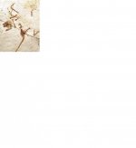
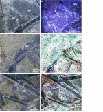
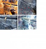
The bizarre scansoriopterygid theropods Yi and Ambopteryx had skin stretched between elongate fingers that form a potential membranous wing. This wing is thought to have been used in aerial locomotion, but this has never been tested. Using laser-stimulated fluorescence imaging, we re-evaluate their anatomy and perform aerodynamic calculations covering flight potential, other wing-based behaviors, and gliding capabilities. We find that Yi and Ambopteryx were likely arboreal, highly unlikely to have any form of powered flight, and had significant deficiencies in flapping-based locomotion and limited gliding abilities. Our results show that Scansoriopterygidae are not models for the early evolution of bird flight, and their structurally distinct wings differed greatly from contemporaneous paravians, supporting multiple independent origins of flight. We propose that Scansoriopterygidae represents a unique but failed flight architecture of non-avialan theropods and that the evolutionary race to capture vertebrate aerial morphospace in the Middle to Late Jurassic was dynamic and complex.
Aerodynamics show membrane-winged theropods were a poor gliding dead-end
iScience advance online publication
doi: 10.1016/j.isci.2020.101574
https://www.cell.com/iscience/fulltext/S2589-0042(20)30766-5
The bizarre scansoriopterygid theropods Yi and Ambopteryx had skin stretched between elongate fingers that form a potential membranous wing. This wing is thought to have been used in aerial locomotion, but this has never been tested. Using laser-stimulated fluorescence imaging, we re-evaluate their anatomy and perform aerodynamic calculations covering flight potential, other wing-based behaviors, and gliding capabilities. We find that Yi and Ambopteryx were likely arboreal, highly unlikely to have any form of powered flight, and had significant deficiencies in flapping-based locomotion and limited gliding abilities. Our results show that Scansoriopterygidae are not models for the early evolution of bird flight, and their structurally distinct wings differed greatly from contemporaneous paravians, supporting multiple independent origins of flight. We propose that Scansoriopterygidae represents a unique but failed flight architecture of non-avialan theropods and that the evolutionary race to capture vertebrate aerial morphospace in the Middle to Late Jurassic was dynamic and complex.