Systematic Paleontology
Order Procellariiformes Fürbringer, 1888
Family
Procellariidae Leach, 1820
Genus
Macronectes Richmond, 1905
Macronectes tinae sp. nov.
ZooBank reg. nr.: urn:lsid:zoobank.org:act:EB59C374-6AE6-4FB8-8D22-0F2747CD6F3A
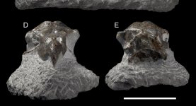
Holotype: NMNZ S.048502 (col. Alastair Johnson, 2017): largely complete skull (Figure 1).
Paratype: NMNZ S.048870 (col. Alastair Johnson, 2019): left humerus, fragmentary, only the shaft and distal end remaining (Figure 3A). The shaft is to be broken near where the crista deltopectoralis would terminate.
Type locality and stratum: New Zealand, North Island, southern Taranaki, H¯awera. Tangahoe Formation. The holotype and paratype were surface collected as beach boulders and do not have an exact Fossil Record Electronic Database number, but see Q21/f0002 for nearby location.
Age: Late Pliocene, Piacenzian (“Waipipian Stage” in the New Zealand scale): constrained to 3.36–3.06 Ma, based on the oxygen isotope stage and magnetic polarity data.
Etymology: The specific epithet honours Tina King, the late partner of fossil collector Alastair Johnson. This giant petrel skull was her favourite fossil, hence the homage.
Measurements: Skull: FTD = 7.2 mm; MFF = 6.9 mm; MIF = 20.5 mm; MUW= 12.6 mm; NL = 23.1 mm; NW = 4.5 mm; PFNW = 22.2 mm; PoW = 48.8 mm (estimate); PrW = 34.9 mm. (estimate); TL = 148 mm. Humerus: total (preserved) length = 166 mm; DW = 26.8 mm; SbW = 14.7 mm; SbD = 6.8 mm.
Diagnosis: Skull: overall smaller size; proportionately longer apertura nasi ossea; apparently shallower os supraocciptale. Humerus: shaft proportionately craniocaudally less deep, with
more delicate appearance than congeners; medial portion of epicondylus ventralis more prominent; fossa medialis brachialis proportionately larger, elongated, and nearly fusiform.
Differential diagnosis: There is little intraspecific and interspecific variation in the skull and humerus morphology between
M. giganteus and
M. halli; barring sexual dimorphism (males are larger
")
and the slightly smaller average size of
M. halli (the size range of the two species completely overlap
. That is to be expected from taxa with little genetic distinction. As such, morphological comparisons can be made between
M. tinae sp. nov. and both living
Macronectes spp. simultaneously.
Skull: The skull of
M. tinae sp. nov. (Figure 1) is smaller than all
Macronectes spp. in the NMNZ collection (Table S1) and can be instantly diagnosed by its size. Barring the size difference, almost all other structures are the same as in living
Macronectes spp. (Figure 2), with two exceptions: the fossa temporalium and the os supraocciptale.
According to the PCA, PC1 explains circa 55% of variance, PC2 26%, and PC3 8% (Table S2). PC1 values are strongly related to almost all measurements, except FTD (distance between fossae temporalium); larger values of PC1 mean larger sizes. PC2 is mostly related to FTD, with larger PC2 values indicating larger FTD. PC3 is related to MFF (minimum interorbital width) and NL (length of the nasal aperture); larger PC3 values indicate larger MFF, but smaller NL. In a PC1 PC2 plot (Figure 5B), there is not much difference between
M. tinae sp. nov. and living
Macronectes spp. (which greatly overlap). However, a PC1 PC3 plot (Figure 5C) shows
M. tinae sp. nov. is separated from the two recent species due to its low PC3 value (potentially due to NL).
The length of the nasal aperture (Table S1) of
M. tinae sp. nov. is roughly the same as in living giant petrels, making it proportionately larger in the fossil (in relation to the rest
of the skull).
Furthermore, the os supraocciptale (supraocciptal bone) of
M. tinae sp. nov. (Figure 1) is apparently shallower than in living
Macronectes spp., even considering the smallest specimens of the latter. The depth of this bone in
M. tinae sp. nov. is likely somewhere between 1/2 and 2/3 of the depth observed in recent
Macronecets spp. (Figure 2) and could be an important diagnostic feature. However, due to the light deformation of the fossil, this cannot be stated with precision, and no reliable measurements could be taken from this bone to include in the PCA. As such, this must remain as a qualitative comparison for the moment.
Finally, the crista nuchalis transversa is apparently more prominent in
M. tinae sp. nov. than in its living congeners, although this feature might have been exacerbated in the
present fossil due to preservation (caudal end of skull lightly crushed; Figure 1).
Humerus: The humerus of
M. tinae sp. nov. is about as big as the smallest Macronectes spp. in the NMNZ collection (e.g., NMNZ OR.015606; Table S1). The distal end of the fossil is more delicate than that of living Macronectes spp., with its shaft being proportionately less deep (Figure 3; Table S1: larger [SbW/SbD] measure in M. tinae sp. nov.). According to
the PCA, PC1 explains 81% of variance and PC2 17% (Table S3). Larger PC1 values mean greater W, SbW, and SbD, while larger PC2 values mean greater SbW and SbD (thicker shaft), but lower W (smaller distal end). By plotting PC1 PC2 (Figure 5A), it is clear that
M. tinae sp. nov. is separated from the two recent species (which largely overlap) due to its different proportions, as explained above.
The base of the processus supracondylaris dorsalis of the fossil has the same shape as found in living Macronectes spp., but its spur-like extension is broken off (Figure 3A). Likewise, the condylus dorsalis (dorsal/external condyle), condylus ventralis (ventral condyle), and epicondylus ventralis (ventral epicondyle) are all worn, but what remains of them has similar shapes to the equivalent condyles found in the living species (Figure 3). The epicondylus ventralis appears to extend further distally than the condylus dorsalis in the fossil (Figure 3A). This may be partly due to damage; nevertheless, it appears that the epicondylus ventralis was more developed in
M. tinae sp. nov. than in its congeners (Table S1: larger [DW/SBW] measure in
M. tinae sp. nov.). Other fulmarine petrel genera tend to have a slightly more prominent epicondylus ventralis than
Macronectes spp. (Figure 4). The fossa medialis brachialis (brachial fossa) of
M. tinae sp. nov. is proportionately larger than in its congeners and has an elongated, nearly fusiform shape (Figure 3A); it is more circular in living
Macronectes spp. (Figure 3B,C). The fossa medialis brachialis in other fulmarine petrel genera is usually similar in shape to that found in modern
Macronectes spp.; however, a few
Thalassoica spp. show a more elongate shape, approaching that seen in
M. tinae sp. nov. (Figure 4). The caudal view of the distal humerus remains obstructed by sediment (Figure 3A).
Fred
Figure 1. Skull (holotype, NMNZ S.048502) of Macronectes tinae sp. nov., partially embedded in matrix, in different views; scale bar = 5 cm. (A) Dorsal view. (B) Lateral view (right). (C) Lateral view (left). (D) Anterior view. (E) Caudal view.