• Welcome to BirdForum, the internet's largest birding community with thousands of members from all over the world. The forums are dedicated to wild birds, birding, binoculars and equipment and all that goes with it.

    Please register for an account to take part in the discussions in the forum, post your pictures in the gallery and more.
ZEISS DTI thermal imaging cameras. For more discoveries at night, and during the day.

Vegaviids from the López de Bertodano Formation (2 Viewers)

albertonykus

Well-known member
de Souza, G.A., B.A. Bulak, M.B. Soares, J.M. Sayão, L.C. Weinschütz, A. Batezelli, and A.W.A. Kellner (2023)
The Cretaceous Neornithine record and new Vegaviidae specimens from the López de Bertodano Formation (Upper Maastrichthian) of Vega Island, Antarctic Peninsula
Anais da Academia Brasileira de Ciências 95: e20230802
doi: 10.1590/0001-3765202320230802

A worldwide revision of the Cretaceous record of Neornithes (crown birds) revealed that unambiguous neornithine taxa are extremely scarce, with only a few showing diagnostic features to be confidently assigned to that group. Here we report two new neornithine specimens from Vega Island (López de Bertodano Formation). The first is a synsacrum (MN 7832-V) that shows a complex pattern of transversal diverticula intercepting the canalis synsacri, as in extant neornithines. Micro-CT scanning revealed a camerate pattern of trabeculae typical of neornithines. It further shows the oldest occurrence of lumbosacral canals in Neornithes, which are related to a balance sensing system acting in the control of walking and perching. The second specimen (MN 7833-V) is a distal portion of a tarsometatarsus sharing with Vegavis iaai a straight apical border of the crista plantaris lateralis. Osteohistologically the tarsometatarsus shows a thick and highly vascularized cortex that lacks any growth marks, resembling Polarornis gregorii. The cortex is osteosclerotic as in other extinct and extant diving neornithines. These new specimens increase the occurrences of the Cretaceous avian material recovered from the Upper Cretaceous strata of the James Ross Sub-Basin, suggesting that a Vegaviidae-dominated avian assemblage was present in the Antarctic Peninsula during the upper Maastrichtian.
 
Systematic Paleontology

Aves Linnaeus 1758
Ornithurae Haeckel 1866
Panneognathae Gauthier & de Queiroz 2001
Vegaviidae Agnolín et al. 2017
Vegavis iaai Clarke et al. 2005
cf. V. iaai

Material
– MN 7832-V, three fused synsacral vertebrae lacking the corpus vertebrae.

Locality and horizon
– Sandwich Bluff, Sandwich Bluff Member, SBM8, 9, 10, 11 or 12 of Roberts et al. (2014), approximately 40 m above the level where Vegavis specimens were recovered, López de Bertodano Formation outcropping in Cape Lamb, Vega Island, Antarctic Peninsula. Upper Maastrichtian (~ 66-68 Mya).

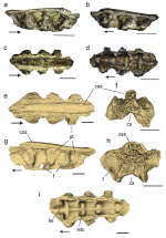
Description and comparisons – The specimen MN 7832-V consists of three fused synsacral neural arches 3.7 mm in length (Fig. 4). It lacks the corpus vertebrae and the most of the processus transversi. Despite its incompleteness, there are no signals of taphonomic deformation. The inner surface of the neural spines has a nacreous aspect, ranging from dusky- to dark-red colors. The proccessus spinosum and ossified tendons are ankylosed into a crista spinosa sinsacri, which is low but exceeds dorsally the processus transversi in lateral view. In Vegavis, the crista spinosa exceeds dorsally the processus transversi in synsacral vertebrae 4th to 8th, similarly to MN 7832-V. The dorsal surface of the crista is straight. The crista decreases in both height and thickness caudally. Two longitudinal sulci extend side by side to the crista. MN 7832-V preserves three proximalmost portions of the processus transversi. Each processus is dorsoventrally tall and mediolaterally short, being laterally rather than caudolaterally or laterocaudally projected. A laterally projected processus also occurs in the preacetabular vertebrae synsacrales in Vegavis (Acosta Hospitaleche & Worthy 2021). In MN 7832-V, the processus transversi lack the end that articulates with the os coxae. The processus transversi are separated by deep concave intervertebral spaces in lateral view. In the floor of these spaces, ventral to each processus transversus lies a series of small foramina intervertebralia. Each intervertebral space bears a small foramen. Similar foramina occur in vertebrae synsacrales 5th to 7th (apparently also in 4th and 9th) of the Vegavis holotype (MLP 93-I-3-1) (Acosta Hospitaleche & Worthy 2021). The smaller size and low density of foramina resemble extant diving birds such as the examined sphenicid Spheniscus magellanicus (MNA 32383, MNA 2858), the suliform Anhinga anhinga (MNA 22863, MNA 26347), rather than cursorial terrestrial Neornithes such as the ratite Rhea americana (MNA 7679) and the charadriid Vanellus chilensis (MNA7004), reinforcing the interpretation of adaptations for a foot-propelled diving ecology for MN 7832-V and Vegavis.
Externally, the contacts between vertebrae are indiscernible. However, the absence of corpus vertebrae exposes, in ventral view, the roof of the canalis synsacri, enabling us to examine the inner structure of the neural arches (Fig. 4d and i). Internally, a sulcus extends longitudinally over the roof of the canalis synsacri, likely accommodating the spinal cord canal. The divisions of the vertebrae are represented by small recesses, identified as lumbosacral transverse canals, that intercept transversally the canal synsacri (Fig. 4i). The functional hypothesis about such canals is that they are part of a secondary balance sensing system, working similarly to the semicircular channels of the inner ear, involved in the control of walking and perching (Stanchak et al. 2020, Jadwiszczak et al. 2022). These canals are found in most neornithine groups (Jalgersma 1951) and, up to now, the oldest record of lumbosacral canals in a synsacrum was reported in a Maastrichthian Ornithurae FMNH PA 741 from Madagascar (O’Connor & Forster 2010). Thus, in the fossil Neornithes context, the specimen MV 7832-V exhibits the oldest record of the lumbosacral canals, since before they were only reported in early Sphenisciformes penguins from the Eocene of Seymour Island, Antarctic Peninsula (Jadwiszczak et al. 2022). The canalis synsacri expands cranially, which likely corresponds to the lumbosacral intumescence (= bulla intumescentia lumbosacralis) that contains the glycogen body (cranium inferior of Barkow 1856). In living birds, this glycogen body is placed medially between two lateral rami of the spinal cord that bifurcate at the level of lumbosacral vertebrae. This feature is present in all crown birds, neornithines, ichthyornithids, and hesperornithids (Acosta Hospitaleche & Worthy 2021). In Vegavis, the lumbosacral intumescence reaches its maximum width around the 6th and 7th vertebrae (Acosta Hospitaleche & Worthy 2021).
Comparison among ornithurines is restricted by the small number of specimens in which the synsacrum is preserved. The crista synsacri of MN 7832-V differs from the ornithurine UA
9601 by having a mediolaterally broader crest, whereas in UA 9601, the crest is sharp. Instead, the dorsal surface of the crista synsacri of MN 7832-V is flattened similar to Apatornis (YPM 1451). The ornithurine UA 9601 lacks the sulci bounding laterally the crista synsacri seen in MN 7832-V and in the anteriormost synsacral vertebrae of Apatornis. In UA 9601, the lumbosacral intumescence occurs around the 4th and 5th vertebrae, whereas in Vegavis and Apatornis it occurs in the 6th and 7th, which we assume to be similar to MN 7832-V. A single large foramen intervertebralis facing laterally lies on the lateral surface of the 4th vertebra in UA 9601 and Apatornis, whereas in Vegavis and MN 7832-V, the foramina occur on the processus transversi of the 5th, 6th, and 7th vertebrae. The morphology of MN 7832-V is consistent with its referral to Vegavis. Based on the morphological comparisons performed here, MN 7832-V is assigned to Vegavis, most likely representing the 5th to 7th vertebra synsacrales. Furthermore, both Vegavis and MN 7832-V possess a pattern of synsacral pneumaticity similar to that of diving birds, reinforcing the hypothesis of a diving ecology for this species.

Fred


Figure 1. Synsacrum MN 7832-V (a-i), photographs of the specimen in (a) right lateral, (b) left lateral, (c) dorsal, and (d) ventral view. Micro CT-scan of MN 7832-V in (e) dorsal, (f) anterior, (g) left lateral, (h) posterior, and (i) ventral views. Scale bar for (a-d) = 10mm, (e-i) 5mm. Arrows indicate anterior side. Abbreviations: cs, canalis synsacri, css, crista spinosa synsacri, f, foramina intervertebralia, lsi, lumbosacral intumescence, lstc, lumbosacral transverse canals, pt, processus transversus.
1702294590743.png
 
Ornithurae Haeckel 1866
Panneognathae Gauthier & de Queiroz 2001
Vegaviidae Agnolín et al. 2017
Polarornis gregorii Chatterjee 1989
cf. Polaronis gregorii

Material
– MN 7833-V, isolated fragment of a tarsometatarsus?

Locality and horizon – Sandwich Bluff, Sandwich Bluff Member, SBM8, 9, 10, 11 or 12 of (sensu Roberts et al. 2014), approximately 40 m above the level where Vegavis specimens were recovered, López de Bertodano Formation outcropping in Cape Lamb, Vega Island, Antarctic Peninsula. Upper Maastrichtian (~66-68 Mya).

Description and comparisons
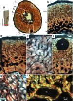
MN 7833-V consists of a small fragment (3.5 mm) of a rod-like bone with a diameter of 8.8 mm, lacking its proximal and distal ends. The specimen was found with no association with the synsacrum MN 7832-V. The incompleteness of the bone precludes its precise identification, but some features are informative. The partial diaphysis has a longitudinal and sharp crest extending along the shaft, but its presumably distal portion is broken. The apical margin of the crest is straight and roughly parallel to the shaft surface. As it extends along the shaft, the crest deflects and gradually merges onto the shaft surface. One of the surfaces of the crest is convex and continuous with the shaft, whereas the opposite surface is concave, which gives to the shaft a half salinon-shape to the transverse section in distal view. Above the crest level, the shaft exhibits a roughly circular cross-section. The combination of straight shaft, roughly circular shape in transversal section, and the presence of a sharp and bowed crest suggests that the specimen represents a hind limb long bone. The preserved shaft is elongated and robust, possessing a thick cortex, similar in proportions to the avian tibiotarsus or tarsometatarsus. In comparison with partially chronocorrelated birds, the crest of MN 7833-V superficially resembles the crista fibularis or tuberositas retinaculi extensoris of the tibiotarsus of Vegavis (Acosta Hospitaleche & Worthy 2021). These crests are sharp and have rounded apical edges as in MN 7833-V. However, they are lower in Vegavis, protruding briefly from the shaft to immediately merge into the bone surface. In addition, the apical margins of these crests are not parallel to the shaft as in MN 7833-V. Instead, the crest of MN 7833-V is more similar to the crista plantaris lateralis (crista dorsalis lateralis of Acosta Hospitaleche & Worthy 2021) of the tarsometatarsus of Vegavis. In both MN 7833-V and Vegavis tarsometatarsus, the crests similarly protrude acquiring a more dorsal inclination. Their apical edges are parallel to the straight shaft. The shafts possess a deeply concave sulcus extensorum on one side, whereas they have a convex shaft in the opposite side. The half ‘salinon-shaped’ cross-section is present in both MN 7833-V and Vegavis. However, unlike Vegavis, MN 7833-V lacks foramina vasculare proximale laterale et proximale on the floor of sulcus extensori. MN 7833-V may represent a fragment from a more distal part of the shaft of the tarsometatarsus than the one preserved in Vegavis, in which the distalmost portion of crista plantaris is missing, preventing us from determining whether the inflection of the crest is similar to MN 7833-V or not.
The anatomy of the crest of MN 7833-V also resembles the crista deltopectoralis of the vegaviids Vegavis, Maaqwi (McLachlan et al. 2017, Acosta Hospitaleche & Worthy 2021), and many extant neornithines such as Gavia adamsii and Haliaeetus leucocephalus (Serrano et al. 2020, Watanabe et al. 2021), with a straight apical margin almost parallel to the shaft. The deflection of the crest within the shaft in lateral view of MN 7833-V approaches angles similar to those of the cited vegaviids. However, MN 7833-V differs from the humerus by having a sinuous apical outline instead straight. Given its morphology, we tentatively identify MN 7833-V as an undetermined vegaviid tarsometatarsus.

Fred


Figure 2. Microstructural pattern of MN 7833-V (a-h), proximal fragment of tarsometatarsus MN 7833-V in lateral view (a), whole cross section of MN 7833-V (b). Two cortical regions showing the large amount of intertrabecular spaces (c) and thick endosteal lamella (d) in different portions of the perimedullary cortex. High-magnification of the cortex showing the clusters of osteocyte lacunae, which characterize the woven-fibered bone matrix (e). Nutrient foramen in subperiosteal cortex (f). High magnification of subperiosteal cortex showing the longitudinal primary osteons (g). High magnification of trabeculae bounded by cement lines in perimedullary cortex (h). Red line in a corresponds to the level where the section was made. The outer cortex face upward in b-g. Scale bar in a is equal to 15 mm. Abbreviations: cl, cement line, el, endosteal lamellae, mc, medullary cavity, nf, nutrient foramen, nvc, neurovascular canals, po, primary osteons, oc, osteocyte clusters, rc, resorption cavities, so, secondary osteon, t, trabeculae, ts, intertrabecular spaces.
1702297088005.png
 
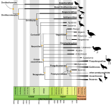
Figure 3. A generalized time‑calibrated phylogeny of Mesozoic birds. The time of divergences of the non-neornithine nodes follows Wang & Lloyd (2016). The split of neornithines in the Early Cretaceous follows Hedges et al. (1996) and Ericson et al. (2006). The positioning of the neornithines is according to Clarke (2004) for Apatornis celer and Iaceornis marshi, McLachlan et al. (2017) and Field et al. (2020) for Vegaviidae, De Pietri et al. (2016) for Teviornis gobiensis. Neogaeornis wetzelli, Limenavis patagonica, Kookne yeutensis, Antarcticavis capelambensis, and Australornis lovei were not submitted to any cladistic analyses. The black horizontal thick lines represent approximated temporal ranges of each taxon. Dotted lines indicate alternative positions of Vegaviidae.
1702297217252.png

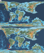
Figure 4. Global paleogeographic maps showing the localities with records of Cretaceous Neornithes. (a) Early Campanian, (b) Late Campanian-Maastrichtian. 1 - Northumberland Formation, British Columbia, Canada (Maaqwi), 2 - Niobrara Formation, Kansas, US (Apatornis and Iaceornis) but see the text to alternative position for these taxa outside Neornithes, 3 - Allen Formation, Rio Negro, Argentina (Limenavis and Lamarqueavis), 4 - Maastricht Formation, Belgium (Asteriornis), 5 - Nemegt Formation, Mongolia (Teviornis, af. Phalacrocoracidae, af. Charadriiformes), 6 - Quiriquina Formation, Chile (Neogaeornis), 7 - Chorrillo Formation, Santa Cruz, Argentina (Kookne, stratigraphic unit ranges from late Campanian to Maastrichtian), 8 - Cape Lamb Member of the Snow Hill Island Formation, Vega Island, Antarctica (Vegavis, Neornithes indet., and Charadriiformes indet.) and López de Bertodano Formation, Seymour Island, Antarctica (Polarornis). Yellow dots mean the Northern Hemisphere neornithines and red dots indicate the Southern Hemisphere neornithines. Plate tectonic maps of Early Campanian (~80 Mya) and Maastrichtian (~70 Mya) by C. R. Scotese (2001), PALEOMAP Project (www.scotese.com).
1702297315331.png

Fred
 

Users who are viewing this thread

Back
Top