• Welcome to BirdForum, the internet's largest birding community with thousands of members from all over the world. The forums are dedicated to wild birds, birding, binoculars and equipment and all that goes with it.

    Please register for an account to take part in the discussions in the forum, post your pictures in the gallery and more.
ZEISS DTI thermal imaging cameras. For more discoveries at night, and during the day.

Zealandornis relictus gen. et sp. nov.; Aegotheles zealandivetus sp. nov. (1 Viewer)

albertonykus

Well-known member
Worthy, T.H., R.P. Scofield, S.W. Salisbury, S.J. Hand, V.L. De Pietri, and M. Archer (2022)
Two new neoavian taxa with contrasting palaeobiogeographical implications from the early Miocene St Bathans Fauna, New Zealand
Journal of Ornithology (advance online publication)
doi: 10.1007/s10336-022-01981-6

Two new neoavian landbirds are reported from the early Miocene St Bathans Fauna from New Zealand. Aegotheles zealandivetus sp. nov. is described from several bones, among which, notably, the tarsometatarsus shows more similarity to New Guinean taxa than to Australian—New Zealand species. Zealandornis relictus gen. et sp. nov. is described from a distal end of a humerus and placed in the new family Zealandornithidae, tentatively among the ‘higher landbirds’ Telluraves, with most similarity to coliiforms. The humerus is of similar size to that of species of Colius and its gracile shaft and very shallow sulcus scapulotricipitalis suggests reduced flying ability. The new species of Aegotheles reinforces the Australasian nature of the Zealandian fauna, while in contrast, Zealandornis relictus gen. et sp. nov. appears to have no close relatives. It is as distinct as Acanthisittidae and Strigopidae among birds, or Leiopelmatidae and Sphenodontidae among the herpetofauna, and like them, represents a similar relictual taxon. Together they confer a highly evolutionarily distinctive nature to the Zealandian fauna concomitant with a minimal 60 million years of isolation.
 
Systematic palaeontology

Class Aves Linnaeus, 1758
Subclass Strisores Baird, 1858, sensu Mayr (2010)
Order Apodiformes Peters, 1940

Material:
NMNZ S.52917 (Fig. 1) has the common avian arrangement of the trochleae metatarsorum II, III and IV directed cranially, which excludes affinity with birds having the zygodactyl condition of the trochlea metatarsi IV rotated plantarly, usually with development of an accessory trochlea (Psittaciformes, Strigiformes, Cuculiformes, Coliiformes, Piciformes (including Picidae, Ramphastidae, Bucconidae, Gabulidae, Indicatoridae), or with those having the heterodactyl condition of trochlea metatarsi II rotated plantarly (Trogoniformes). Tarsometatarsi of bucerotiforms differ markedly from the fossil in having the trochleae essentially in one plane, fossa metatarsi I deeply incising the medial margin of the shaft, trochlea metatarsi II markedly prominent medially and trochlea metatarsi III short. Tarsometatarsi of all passerines differ markedly from the fossil in that the trochleae are in one dorsoplantar plane and have near equal distal extent.
Tarsometatarsi of procellariiforms, specifically species of Pelecanoides, which is represented by P. miokuaka Worthy et al., 2007 in the St Bathans Fauna (Worthy et al. 2007), differ greatly as follows: they have no, or a very weakly formed, fossa metatarsi 1; the foramen vasculare distale is larger and preceded by a deep sulcus; the trochlea metatarsi II is distinctly shorter than the trochlea metatarsi IV and is deeply grooved; and the trochlea metatarsi IV is grooved dorsally.
The fossil shows closest similarity to the tarsometatarsi of some caprimulgiforms, e.g., Eurostopodus argus and aegothelids, but those of species of Eurostopodus differ notably in having the proximal facet in the fossa metatarsi I prominent medially, cristae plantares medialis et lateralis more robust, distally shorter trochleae that diverge symmetrically from the shaft and are more arched so that, plantarly, the gap separating trochleae metatarsorum II and IV is less than the width of trochlea metatarsi III, and trochleae metatarsorum III and IV have shallow grooves dorsally, distally and plantarly. The tarsometatarsi of aegothelids have most similarity to the fossil.
 
Family Aegothelidae Bonaparte, 1853
Genus Aegotheles Vigors & Horsfield, 1827
Aegotheles zealandivetus sp. nov.
urn:lsid:zoobank.org:act:89951097-E888-4589-9FD2-8E928B40FAF0.

Holotype: NMNZ S.52917, a distal right tarsometatarsus, with part of the shaft and three trochleae exhibiting slight wear but otherwise well-preserved (Fig. 1A–E).

Etymology: From Zealandia, the continent on which New Zealand is emergent and vetus Latin adjective for old, ancient, or former, to reflect the fact that the new species was an old inhabitant of Zealandia, masculine.

Type locality: Bed HH1b, c.10 cm thick sand and coarse cobble layer, 9.5–9.58 m above base of Bannockburn Formation, Trench excavation, foot of hill 50 m across terrace from riverbank at 44.54.493°S; 169.85844°E; Manuherikia River, Home Hills Station, Otago, New Zealand. NZ Fossil Record File Number H41/f0103.

Stratigraphy and age: Bannockburn Formation, Manuherikia Group, early Miocene (19–16 Ma).

Diagnosis: The new taxon is an aegothelid having the following unique combination of characters for the distal tarsometatarsus: The fossa metatarsi I is deep, broad, not impacting on the medial profile of the shaft; the foramen vasculare distale is small, close to the incisura lateralis, and preceded by a short shallow sulcus; the trochleae diverge asymmetrically; the trochlea metatarsi II is directed somewhat medially making the medial profile concave; the trochlea metatarsi IV is slightly offset from the shaft, directed distally, resulting in convex lateral profile; the dorsal surface proximally adjacent to trochlea metatarsi III is flat, lacking a sulcus extending proximally from the incisura medialis; the distal projection of trochlea metatarsi II is slightly less than that of trochlea metatarsi IV, with 25% of the length of trochlea metatarsi III exceeding both; the trochlea metatarsi II lacks a groove dorsally; the trochlea metatarsi IV has a shallow groove distally but none dorsally; and the trochlea metatarsi III is deeply grooved dorsally, distally and plantarly, deeper than wide, with rims parallel in plantar aspect.

Differential diagnosis: Differs from Aegotheles savesi, A. novaezealandiae, A. salvadorii and A. cristatus on account of the following features: the dorsal surface proximal to trochlea metatarsi III is flat and merges with an evenly convex facies extending to the medial margin proximal to the trochlea metatarsi II without interruption by a groove extending from the intertrochlear incision (as opposed to being convex with a deep groove); the foramen vasculare distale is smaller; the trochlea metatarsi II is shorter than the trochlea metatarsi IV (as opposed to a relatively longer trochlea metatarsi II that distally exceeds trochlea metatarsi IV, or which may distally exceed trochlea metatarsi III in A. novaezealandiae).
Differs from A. novaezealandiae on account of: the trochlea metatarsi IV extends distally slightly past the trochlea metatarsi II; the trochlea metatarsi IV projects distally straight (not directed laterally); the fossa metatarsi I is larger and more excavated.
Differs from A. savesi on account of: the dorsal surface is flatter; the trochlea metatarsi II does not equal or surpass distally the trochlea metatarsi IV in medial/dorsal views; the trochlea metatarsi II is more plantarly retracted; the projection on the medial rim of the trochlea metatarsi II is smaller, even allowing for damage (A. savesi has a robust plantar flange); the trochlea metatarsi II is broader mediolaterally (dorsal view); the rims of the trochleae are more pronounced; there is a deeper furrow on trochlea metatarsi III, with rims that are parallel and not tapered in plantar aspect (A. savesi has proximally convergent rims); the trochlea metatarsi IV has a groove distally (lacking) and its outer rim lacks plantar projection (in A. savesi, the outer rim has a narrow flange extending about 30% of its depth plantar to the inner rim); the foramen vasculare distale is round, smaller and situated closer to incisura intertrochlearis; the fossa metatarsi I is wider/broader; and the cristae plantares medialis et lateralis is stronger.
Differs from Aegotheles cristatus and A. salvadorii on account of: larger size (Table 1); the trochlea metatarsi II is not as medially splayed; the trochlea metatarsi IV is directed mainly distally (more laterally) and is more laterally positioned relative to the shaft so that the profile is convex (versus straight in A. cristatus and A. salvadorii); the trochlea metatarsi III is deeply grooved; the trochlea metatarsi IV is shallowly grooved distally (in A. cristatus and A. salvadorii, all trochleae lack a groove both dorsally and distally).
Most similar to those of A. crinifrons and A. insignis, especially in the minimal development of a sulcus and ridge that extends proximally to the incisura medialis and the trochlea metatarsi III, respectively, and in the trochlea metatarsi II being comparatively short. Differs from A. crinifrons on account of: the shaft is more slender; the trochlea metatarsi II is shorter and level with the trochlea metatarsi IV; the trochleae metatarsorum III et IV are grooved; the trochlea metatarsi III has rims that are parallel in plantar view (convergent proximally). Differs from A. insignis on account of: the shaft is more slender; the trochlea metatarsi II is longer (IV much exceeds II in insignis); the trochlea metatarsi III is deeply grooved dorsally (lacking); the foramen vasculare distale is relatively larger.
Aegotheles tatei is the only similar-sized aegothelid that the specimen was not compared with, but it was considered conspecific with A. insignis until recently, so is probably very similar to that taxon; moreover, all these modern birds are separated from the fossil taxon by a minimum of 16 million years so are unlikely to be conspecific.

Referred specimens: All from beds on the East bank of the Manuherikia River, Home Hills Station, St Bathans, Otago, NZ. NMNZ S.42800, a proximal right tarsometatarsus, Bed HH1a, 6.88–7.0 m above base of Bannockburn Formation, at
44.907944S, 169.858222E. NZ Fossil Record File Number H41/f88. (Schwarzhans et al. 2012; Worthy et al. 2022). CM 2013.18.1127, cranial part of a right coracoid (Fig. 2A–C), Bed HH2, 21.02–21.48 m above the base of the Bannockburn Formation, at 44.907861°S 169.857389°E; NZ Fossil Record File Number H41/f87. NMNZ S.51974, proximal right ulna (Fig. 2G), Bed HH1b, details as per type locality. NMNZ S.52761, distal right tibiotarsus (Fig. 2J), Bed HH1b, details as per type locality.

Measurements (mm): Holotype, NMNZ S.52917, distal right tarsometatarsus, preserved length 12.0; shaft width 2.1; shaft depth 1.8; distal width 5.1. NMNZ S.42800, proximal right tarsometatarsus: preserved length 11.0; proximal width 4.3; proximal depth 4.1 mm; least shaft width 1.6 mm. NMNZ S.51974, proximal right ulna: maximum proximal width 4.5. CM 2013.18.1127, cranial part right coracoid: preserved length 12.6; cotyla scapularis to proximal side facies articularis humeralis 3.5. NMNZ S.52761, distal right tibiotarsus: distal width 4.4; proximodistal length condylus lateralis 3.1; proximodistal length condylus medialis 2.9; depth condylus medialis 4.1. See Table 1.


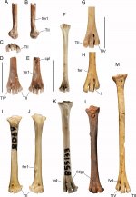
Fig. 1 Tarsometatarsi of species of Aegotheles. Aegotheles zealandivetus sp. nov. NMNZ S.52917, distal right tarsometatarsus (A–E) in lateral (A), medial (B), distal (C),
dorsal (D), plantar (E), aspects; Aegotheles cristatus SAM B.55133 (F, K); Aegotheles
novaezealandiae
CM Av16996 (holotype), left tarsometatarsus (reversed) (H, M); A. novaezealandiae CM Av17503 (paratype), distal left tarsometatarsus (reversed) (G). A. insignis YPM ORN 104654 (I, J); A. savesi right tarsometatarsus, (IANCP unregistered: 100–110 cm Testpit 3, Pindai, New Caledonia) (L). Abbreviations: cpl,
crista plantaris lateralis; fm1, fossa metatarsi I; fvd, foramen vasculare distale; il, incisura intertrochlearis lateralis; TII, trochlea metatarsi II; TIV, rochlea metatarsi IV. Scale bars are 10 mm, and apply to images A–H. Images I–M are scaled so the distal width is the same as in (D, E)

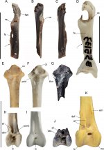
Fig. 2 Bones of species of Aegotheles: coracoids (A–D); ulnae (E–G); and tibiotarsi (H–J). Aegotheles zealandivetus sp. nov. cranial part right coracoid CM 2013.18.1127 (A–C), proximal right ulna NMNZ S.51974 (G), distal right tibiotarsus NMNZ S.52761 (J); Aegotheles cristatus SAM B.55133 (D, F, H), A. novaezealandiae CM Av28220 (paratype), proximal left ulna [reversed] (E), CM Av18240, distal left tibiotarsus reversed (K); A. insignis YPM ORN 104654, surface scan of specimen (I). Abreviations: cs, cotyla scapularis; cv, cotyla ventralis; dsf, distal sulcus of musculus fibularis; el, tuberositas retinaculi extensoris lateralis; em, tuberositas retinaculi extensoris medialis; fah, facies articularis humeralis; fs, foramen nervi supracoracoidei; ic, incisura intercondylaris; lc, condylus lateralis; mc, condylus nedialis; ol, olecranon; pcd, processus cotyla dorsalis; tlcv, tuberculum ligamentum collateralis ventralis; arrow points distal end of the processus procoracoideus. Scale
bars = 10 mm
 

Attachments

  • Aegotheles zealandivetus.jpg
    Aegotheles zealandivetus.jpg
    375.8 KB · Views: 0
  • Aegotheles zealandivetus - 2.jpg
    Aegotheles zealandivetus - 2.jpg
    483.9 KB · Views: 0
Last edited:
Order incertae sedis.
Family Zealandornithidae fam. nov.
urn:lsid:zoobank.org:act:EE0CDC0E-A6EB-4118-96FE-9F8C59D3CC09.

Included taxa: Zealandornis relictus gen. et sp. nov.

Diagnosis: A small coly-like bird, with distal humeri characterised by the following unique combination of characters: a large, robust, dorsally prominent processus supracondylaris dorsalis with its tip located just proximal to the condylus dorsalis; the scar the for palmar branch of musculus extensor carpi radialis is located ventrally adjacent to the processus supracondylaris dorsalis, proximally enclosing a deep lenticular fossa adjacent to the condylus dorsalis; a deep fossa brachialis, restricted to the ventral half of the bone, extending proximal to the processus supracondylaris dorsalis, and narrowly separated from the ventral margin; an undeveloped epicondylus dorsalis, not projected dorsal to the condylus dorsalis; the condylus dorsalis is widely separated from the tuberculum supracondylare ventrale; the condylus ventralis is globose, distally prominent; a robust ventrally prominent processus flexorius–tuberculum supracondylare ventrale complex that lacks a prominent epicondylus ventralis; a distally short processus flexorius, greatly surpassed by the condylus ventralis; no distinct sulcus scapulotricipitalis (autapomorphy); the tuberculum supracondylare ventrale is robust, cranially elevated, lacking a distinct facet for the attachment of ligamentum collaterale ventrale (autapomorphy), and flattened ventrally.
The overall form, with maximum distal width about twice as wide as the proximodistal length from the condylus dorsalis to the processus supracondylaris dorsalis, the processus supracondylaris dorsalis being large and prominent, and the processus flexorius–tuberculum supracondylare ventrale complex being strongly ventrally prominent, is consistent with it deriving from a bird in one of the following neoavian ordinal lineages, Cuculiformes, Strigiformes, Caprimulgiformes, and Coraciimorphae, including Coliiformes, Leptosomatiformes, Trogoniformes, Bucerotiformes, Piciformes, Coraciiformes, and Passeriformes. It is distinguished from all these taxa by lack of a well-marked facet on the tuberculum supracondylare ventrale, and from all except podargids by a lack of a distinct sulcus scapulotricipitalis.
It is further distinguished from passeriforms by the dorsal location of the scar for the palmar branch of musculus extensor carpi radialis; from strigiforms, by the more proximal location of the processus supracondylaris dorsalis, a distally shorter processus flexorius, lack of ventral prominence of the epicondylus ventralis and smaller size; from cuculids by the processus supracondylaris dorsalis being less offset proximally from the condylus dorsalis, the scar for the palmar branch of musculus extensor carpi radialis more dorsoventrally elongate, less ventrally prominent processus flexorius–epicondylus ventralis complex, distally shorter processus flexorius, more elongate fossa musculi brachialis extending proximal to the processus supracondylaris dorsalis; from caprimulgiforms, by the more proximal location of the processus supracondylaris dorsalis, more globose condylus ventralis, and a wider tuberculum supracondylare ventrale that is more widely separated from the condylus dorsalis; further differentiated from podargids, by a larger more prominent processus supracondylaris dorsalis, a better defined lenticular sulcus proximal to condylus dorsalis; from caprimulgids, by a much larger more prominent processus supracondylaris dorsalis, where the palmar branch of musculus extensor carpi radialis is ventrally adjacent to the scar for the dorsal branch, not offset proximally from it; from aegothelids, by the lack of a dorsally prominent epicondylus dorsalis, the presence of a robust dorsally prominent processus supracondylaris dorsalis, and the lack of a ventrally projecting triangular epicondylus ventralis; from all trogoniforms and bucerotiforms, in having a robust dorsally prominent processus supracondylaris dorsalis; from piciforms, by having a distally short processus flexorius rather than one that is elongate and surpasses the condylus ventralis, and from ramphastids among piciforms, by the large dorsally prominent processus supracondylaris dorsalis; from all coraciiforms, by a globular condylus ventralis, lack of a distinct ventrally prominent epicondylus ventralis, and lack of a dorsally prominent epicondylus dorsalis; and from coliiforms, by a distally shorter processus flexorius, lack of projection for the epicondylus ventralis, and less separation of fossa musculi brachialis from the lenticular fossa proximal to the condylus dorsalis.
The lack of a distinct sulcus scapulotricipitalis confers superficial similarity to palaeognaths—lithornithids and tinamids—although this feature is also seen in many galliforms and podargids. Both lithornithids and tinamids differ by a much narrower gap between the condylus dorsalis and the tuberculum supracondylare ventrale (<tuberculum width), smaller processus supracondylaris dorsalis, a distinct and large fossa for insertion of the proximal head of musculus pronator superficialis, a stouter shaft, and much larger size. Lithornithids differ further by the fossa musculi brachialis extending to the dorsal margin and a far more distally elongate processus flexorius (Houde 1988). All compared tinamids, and see Bertelli et al. (2014), have a shallow fossa musculi brachialis of variable extent and a much smaller and not proximally hooked processus supracondylaris dorsalis. These features make it very unlikely that the fossil could be associated with Proapteryx micromeros Worthy TH, Worthy JP, Tennyson, Salisbury, Hand, Scofield, 2013, a small kiwi known from the St Bathans fauna (Worthy et al. 2013a, b). However, we explored the relative size of NMNZ S.52077 and P. micromeros, wherein a ratio of the minimum femur shaft width to distal width of the humerus of 0.66 is much higher than that for volant tinamous and very much less than that for Apteryx owenii (see SI); the distal humerus of all species of Apteryx have amorphous shapes, wherein no typical humeral morphology is present. While the proportions are consistent with a slight relative size reduction in a humerus for the given size of the femur of Proapteryx, the major differences in morphology from volant palaeognaths precludes this association.

Zealandornis relictus gen. et. sp. nov. (Fig. 3A–D).
urn:lsid:zoobank.org:act:B36C310E-FCCB-4E7C-BE75-007911B6676B.
urn:lsid:zoobank.org:act:B733FC16-ACFA-4771-BB06-0586CDF606C3.

Holotype: NMNZ S.52077, distal right humerus (Fig. 3A–D), collected on 15 January 2008 by a University of New South Wales led (S. Hand, M. Archer, et al.) expedition, jointly involving Adelaide University of South Australia (T.H. Worthy et al.), the Museum of New Zealand (A. Tennyson) and the Canterbury Museum (R.P. Scofield).

Diagnosis: As for family.

Type locality: Bed HH1a, Manuherikia River, 6.88–7.0 m above the base of the Bannockburn Formation, East bank Manuherikia River, Home Hills Station, St Bathans, Otago, NZ at 44.907944°S, 169.858222°E. NZ Fossil Record File Number H41/f88. (Schwarzhans et al. 2012; Worthy et al. 2022).

Stratigraphy and age: Lower Bannockburn Formation, Manuherikia Group, early Miocene (19–16 Ma).

Etymology: The genus name captures the origin of this taxon from Zealandia, gender masculine, with the specific epithet to reflect one remaining or left behind.

Measurements (mm): Maximum distal width, processus supracondylaris dorsalis–epicondylus ventralis, 5.4; distal width condylus dorsalis–epicondylus ventralis, 5.0; length processus supracondylaris dorsalis–distal margin condylus dorsalis, 2.9; minimum shaft diameter as preserved, 2.0.

Fred


Fig. 3 Distal right humeri. Zealandornis relictus gen. et sp. nov. NMNZ S.52077 in cranial (A), caudal (B), dorsal (C), and ventral (D) aspect. Aegotheles cristatus SAM B.55133 (E); Colius striatus USNM 558545 (F); Cacomantis flabelliformis SAM B.55124 (G). Abbreviations: cd, condylus dorsalis; cv, condylus ventralis; ed, epicondylus dorsalis; ev, epicondylus ventralis; fb, fossa musculi brachialis; flcv, facet for the attachment of ligamentum collaterale ventrale on the tuberculum supracondylare ventrale; ls, lenticular sulcus; pbmr, scar for the palmar branch of m. extensor carpi radialis; pf, processus flexorius; psd, processus supracondylaris dorsalis; sh, sulcus humerotricipitalis; tsv, tuberculum supracondylare ventrale. Scale bar = 5 mm
 

Attachments

  • Zealandornis relictus.jpg
    Zealandornis relictus.jpg
    284.2 KB · Views: 0
Last edited:
Warning! This thread is more than 2 years ago old.
It's likely that no further discussion is required, in which case we recommend starting a new thread. If however you feel your response is required you can still do so.

Users who are viewing this thread

Back
Top