• Welcome to BirdForum, the internet's largest birding community with thousands of members from all over the world. The forums are dedicated to wild birds, birding, binoculars and equipment and all that goes with it.

    Please register for an account to take part in the discussions in the forum, post your pictures in the gallery and more.
ZEISS DTI thermal imaging cameras. For more discoveries at night, and during the day.

‘Corvus fangshannus’ Hou, 1982, a junior synonym of Corvus corax Linnaeus, 1758 (1 Viewer)

Fred Ruhe

Well-known member
Netherlands
Thomas A. Stidham, Jingmai O'Connor & Zhiheng Li, 2023

The Pleistocene Zhoukoudian ‘Peking Man’ site records the first Beijing (China) evidence of the Northern Raven (Corvus corax)

Journal of Ornithology

Abstract: The Pleistocene Zhoukoudian ‘Peking Man’ site records the first Beijing (China) evidence of the Northern Raven (Corvus corax) - Journal of Ornithology

Study of the holotype and only known material of the purportedly extinct corvid species ‘Corvus fangshannus’ from the late Middle or Late Pleistocene locality 3 of the UNESCO Zhoukoudian ‘Peking Man’ site in Beijing, China, documents its identification instead as a member of the sedentary Northern Raven (Corvus corax) lineage. Shared features of the humerus including those consistent with species of Corvus (protruding ventral tubercle and rounded and not prominent bicipital crest) and a combination of features (including overall size and the presence of a concave ventral margin where the ventral supracondylar tubercle projects ventral to the diaphysis) help to support the allocation of this Pleistocene material to that particular species’ lineage. Ravens do not occur historically within the Beijing Municipality, and this reidentified fossil is the first record from the area. When considered alongside a similar extralimital record based on a recently reported Middle Pleistocene raven skull from Liaoning Province, these fossils together may indicate that the Northern Raven’s prehistoric geographic distribution in northeastern China encompassed a broader area south of its current distribution extending to ~ 40–41 degrees latitude. Changes to the raven’s geographic distribution likely are linked to shifts between cooler and drier climates and warmer ones, along with their interactions with extinct non-analogous vertebrate communities of the Pleistocene. These ravens would have scavenged alongside hyenas, other large mammalian carnivores, and even early humans in northern China.

Enjoy,

Fred
 
Last edited:
Systematic Paleontology

Corvidae
Vigors, 1825.
Corvus Linnaeus, 1758.
Corvus corax Linnaeus, 1758.

Junior synonym: Corvus fangshannus Hou (1982) from Locality 3 of Zhoukoudian, Beijing, China.

Newly referred fossil material: IVPP V6437 (holotype left humerus and left tibiotarsus of ‘C. fangshannus’) from Locality 3 of Zhoukoudian, Beijing, China.

Description of the holotype material of ‘C. fangshannus’

We provide a description of the holotype material to help communicate the morphological features present that help to identify the specimen as a member of the C. corax group and because an extensive description has never been published for this material. The holotype consists of a left humerus and a left tibiotarsus that may or may not belong to the same individual. The humerus is mostly complete except for a few small missing chips of bone and small perforations on the deltopectoral crest, diaphysis, and on the distal end associated with a crack through the specimen (Fig. 2). The humerus has a total length of 86.9 mm, a distal dorsoventral width of 19.6 mm, and proximal dorsoventral width of 24.5 mm. The transverse ligament groove is a broad concave area rather than a narrow groove. Dorsal to it and adjacent (distal) to the humeral head and dorsal tubercle, the cranial surface also is concave and separate from the ventrally positioned groove. The humeral head does not form a lip or overhang over the capital incisure or humeral shaft. The long axis through the ventral tubercle is oblique to the humeral long axis with the proximal end ventral to the distal end. The bicipital crest and ventral rim of the pneumotricipital fossa are thickened. There is a single pneumatic opening within the single pneumotricipital fossa, and it is subcircular in outline positioned near the dorsal crus. The ventral wall of the pneumotricipital fossa is damaged and there may have been some much smaller pneumatic foramina or perforations within the fossa. The bicipital crest has an overall straight margin (cranial/caudal view) and smoothly curves into the diaphysis distally. The dorsal surface of the deltopectoral crest is concave, and the crest extends distal to the bicipital crest. The dorsal tubercle faces dorsoproximally and is positioned dorsal to the humeral head on the proximal surface of the bone. The m. brachialis fossa is elongate and positioned along the ventral edge of the diaphysis (Fig. 2). It narrows to a point proximally. On the ventral face of the bone just distal to the m. brachialis fossa is a muscular origination pit for one of the pronator muscles, and that pit is just proximal to the facet for the ventral collateral ligament on the ventral supracondylar tubercle. That facet is subcircular in outline and is directed craniodistally. The ventral surface of the humerus adjacent to the ligament facet is slightly concave. There is a small tubercle located proximal to the notch between the dorsal and ventral condyles. The dorsal condyle is fractured and partially missing from a crack through the distal end. The dorsal supracondylar process is large and projects proximally. The process is bifurcated with a large bulbous cranial branch and a more caudally positioned and smaller branch that has been broken away. There is a concave facet on the caudal face of the dorsal supracondylar process. The olecranon fossa is deeply inset with a concave area extending proximally into the distal face of the humerus dorsal to the dorsal condyle. There is a m. scapulotricipitalis fossa bounded dorsally by a broad convex area and ventrally by an elongate narrow ridge that extends proximally to near the level of the proximal tip of the dorsal supracondylar process. The flexor process is large, positioned caudal to the condyles, and has a large facet for muscle origination on its caudodistal face. The left tibiotarsus is missing much of its proximal articular surface, adjacent cnemial crests, and the distal approximately half of both distal condyles (Fig. 3). Its preserved length is 100.0 mm with a distal width across the condyles of 10.9 mm. There are some missing fragments of bone associated with fractures in the diaphysis. In its current state, the distal end appears to be rotated relative to the proximal part with the condyles directed laterally (Fig. 3), and that condition contrasts with the drawing provided by Hou (1993, Fig. 71c). That rotation may suggest that the bone was reconstructed missing some of the shaft (and hence the bone was longer than its current state) because the remaining fragments appear to fit one another for the most part. The medial side of the proximal articular surface is preserved. Only the bases of the cnemial crests are present, but the preserved portions show that they are proximodistally short. The fibular crest is prominent and thick with its distal end positioned slightly lateral to its proximal end. The thin crestr low ridge that extends distal from the cranial cnemial crest ends near the level of the proximodistal midpoint of the fibular crest. The extensor sulcus is oriented somewhat oblique to the diaphysis with its proximal preserved end in the medial part of the shaft, ending at a fracture, and the more distal end at the supratendinal bridge located more laterally (Fig. 3). The elongate attachment for the medial side of the extensor retinaculum lies proximal to the level of the m. fibularis groove on the lateral side of the shaft. The length of the m. fibularis groove is about the same length as that of the supratendinal bridge. The groove’s medial edge is formed by the lateral attachment site of the extensor retinaculum (which extends onto the supratendinal bridge), and it has a lateral flange of bone that extends laterally beyond the tibia’s shaft. The distal opening of the extensor groove beyond the ossified bridge is directed distomedially. The lateral condyle extends proximal to the medial condyle and the intercondylar sulcus is a wide U-shape (distal view) with the deepest part located medial to its mediolateral midpoint. There is a small lateral epicondyle preserved. Most of the medial side of the medial condyle is missing. Only the proximal end of the articular surface for the tibial cartilage is present. The medial condyle appears to be positioned medial to the tibial shaft.

Fred


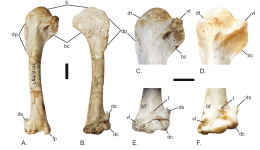
Fig. 1 Comparison of the fossil humerus IVPP 6437, newly referred to Corvus corax (holotype of ‘Corvus fangshannus’), with that from a modern Corvus corax FMNH 465346. Humerus IVPP 6437 in A. caudal view and B. cranial view. Proximal humerus of C. IVPP 6437 and D. FMNH 465346 in caudal view. Distal humerus of E. IVPP 6437 and F. FMNH 465346 in cranial view. Note the dashed line
indicates one branch of the broken bifurcation of the dorsal supracondylar process.
Scale bars equal 1 cm.
Osteological abbreviations: bc—bicipital crest; bf—m. brachialis fossa; dc—dorsal condyle; dp—
deltopectoral crest; ds—dorsal supracondylar process; dt—dorsal tubercle; fp—flexor process; h—humeral head; t—tubercle; vl—facet for the ventral collateral ligament; and vt—ventral tubercle

1693381955581.png
 

Users who are viewing this thread

Back
Top