Fred Ruhe
Well-known member

Gerald Mayr, Ursula Göhlich, Zbynec Roček, Alfred Lemierre, Viola Winkles & Georgios L. Georgalis, 2023
Reinterpretation of tuberculate cervical vertebrae of Eocene birds as an exceptional anti-predator adaptation against the mammalian craniocervical killing bite
Journal of Anatomy.
doi:10.1111/joa.13980. PMID 37990985
Abstract: Reinterpretation of tuberculate cervical vertebrae of Eocene birds as an exceptional anti-predator adaptation against the mammalian craniocervical killing bite - PubMed
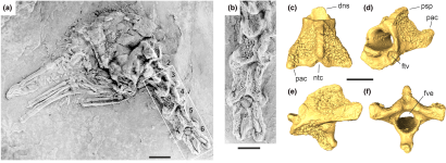
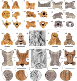
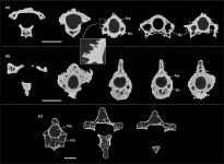
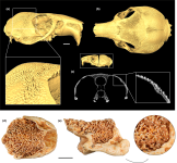
We report avian cervical vertebrae from the Quercy fissure fillings in France, which are densely covered with villi-like tubercles. Two of these vertebrae stem from a late Eocene site, another lacks exact stratigraphic data. Similar cervical vertebrae occur in avian species from Eocene fossils sites in Germany and the United Kingdom, but the new fossils are the only three-dimensionally preserved vertebrae with pronounced surface sculpturing. So far, the evolutionary significance of this highly bizarre morphology, which is unknown from extant birds, remained elusive, and even a pathological origin was considered. We note the occurrence of similar structures on the skull of the extant African rodent Lophiomys and detail that the tubercles represent true osteological features and characterize a distinctive clade of Eocene birds (Perplexicervicidae). Micro-computed tomography (μCT) shows the tubercles to be associated with osteosclerosis of the cervical vertebrae, which have a very thick cortex and much fewer trabecles and pneumatic spaces than the cervicals of most extant birds aside from some specialized divers. This unusual morphology is likely to have served for strengthening the vertebral spine in the neck region, and we hypothesize that it represents an anti-predator adaptation against the craniocervical killing bite ("neck bite") that evolved in some groups of mammalian predators. Tuberculate vertebrae are only known from the Eocene of Central Europe, which featured a low predation pressure on birds during that geological epoch, as is evidenced by high numbers of flightless avian species. Strengthening of the cranialmost neck vertebrae would have mitigated attacks by smaller predators with weak bite forces, and we interpret these vertebral specializations as the first evidence of "internal bony armor" in birds.
Enjot,
Fred
Last edited:


